Molecular Imaging

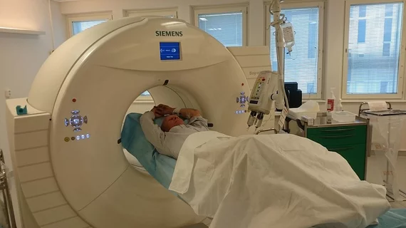
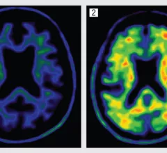
Molecular imaging (also called nuclear medicine or nuclear imaging) can image the function of cells inside the body at the molecular level. This includes the imaging modalities of positron emission computed tomography (PET) and single photon emission computed tomography (SPECT) imaging. How does PET and SPECT imaging work? Small amounts of radioactive material (radiopharmaceuticals) injected into a patient. These can use sugars or chemical traits to bond to specific cells. The radioactive material is taken up by cells that consume the sugars. The radiation emitted from inside the body is detected by photon detectors outside the body. Computers take the data to assemble images of the radiation emissions. Nuclear images may appear fuzzy or ghostly rather than the sharper resolution from MRI and CT.  But, it provides metabolic information at a cellular level, showing if there are defects in the function of the heart, areas of very high metabolic activity associated with cancer cells, or areas of inflammation, data not available from other modalities. These noninvasive imaging exams are used to diagnose cancer, heart disease, Alzheimer’s and Parkinson’s disease, bone disorders and other disorders. 

Novel agent emerges for early Parkinson’s

A new imaging agent is showing potential for the early detection of Parkinson’s disease (PD) by identifying a functional loss of dopamine neurons, according to a study published online Nov. 28 by the Journal of Nuclear Medicine.

Molecular imaging device market expected to rise to $3B by 2018

The current global market for molecular imaging technology is estimated at $2.2 billion this year and is expected to increase at a compound annual growth rate of 6.2 percent, according to a November 2013 market research report from BCC Research.

Thumbnail

Estimated Alzheimer's spikes to 135 million by 2050

New data regarding the prevalence of Alzheimer's disease is looming far and above previous estimates: 17 percent higher than previously thought, according to statistics from Alzheimer's Disease International.

RSNA: Amyloid studies ‘disappointing’

CHICAGO--The quest for a clear way forward in neurodegenerative imaging and therapeutics continues, with many of the same challenges continuing from previous years, according to a neuroimaging symposium during the Radiological Society of North America’s (RSNA) 99th annual meeting.

Dose optimization technologies at RSNA

A number of exhibitors in attendance at last week’s Radiological Society of North America (RSNA) annual meeting in Chicago presented milestones in dose-technology development and hospital integration.

More PET/MR presence at this year’s RSNA

All major manufacturers of molecular imaging technologies now have PET/MR systems in their product portfolios. While Siemens is the only company to have an FDA approved, simultaneous PET/MR system on the market, a number of reports indicate that GE Healthcare is not too far behind in the research and development phase of an on-board and fully integrated PET/MR scanner, as evidence would have it at the 2013 Radiological Society of North America (RSNA) annual meeting still underway in Chicago until its conclusion tomorrow.

Thumbnail

RSNA: Philips unveils advanced PET/CT detector

CHICAGO—Philips presented the new Vereos PET/CT system with solid state detectors at the 99th Radiological Society of North America (RSNA) annual meeting held Dec. 1-6 at McCormick Place convention center.

Ru-82 3D PET/CT MPI training leads to 94% inter-reader agreement

The rubidium-ARMI (alternative radiopharmaceutical for myocardial imaging) multicenter trial has established a bar for assessing myocardial perfusion imaging (MPI) that meets or even exceeds previous SPECT MPI reader scores after thorough consensus review, according to a study published Nov. 18 in the Journal of Nuclear Medicine.