Molecular Imaging

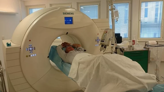
Molecular imaging (also called nuclear medicine or nuclear imaging) can image the function of cells inside the body at the molecular level. This includes the imaging modalities of positron emission computed tomography (PET) and single photon emission computed tomography (SPECT) imaging. How does PET and SPECT imaging work? Small amounts of radioactive material (radiopharmaceuticals) injected into a patient. These can use sugars or chemical traits to bond to specific cells. The radioactive material is taken up by cells that consume the sugars. The radiation emitted from inside the body is detected by photon detectors outside the body. Computers take the data to assemble images of the radiation emissions. Nuclear images may appear fuzzy or ghostly rather than the sharper resolution from MRI and CT.  But, it provides metabolic information at a cellular level, showing if there are defects in the function of the heart, areas of very high metabolic activity associated with cancer cells, or areas of inflammation, data not available from other modalities. These noninvasive imaging exams are used to diagnose cancer, heart disease, Alzheimer’s and Parkinson’s disease, bone disorders and other disorders. 

Radiogenomics: A closer look

The radiologic and pathologic methods of the past century are being replaced by recent knowledge about disease pathology and genetic specialty, moving away from the appearance of disease toward the expression of genes that play an important role in risk stratification, diagnostics, monitoring disease progression and projecting survival.

Alzheimer’s birthplace in the brain announced

The lateral entorhinal cortex has been named the area in the brain where neurodegenerative disease originates, providing new information about how neuronal dysfunction leads to Alzheimer’s progression, according to a study published Dec. 22 in Nature Neuroscience.

Thumbnail

HIV takes a hit with radioimmunotherapy

Targeted radionuclide therapy could change the whole paradigm used to treat human immunodeficiency virus (HIV), according to one of the most remarkable studies to emerge from the recent Radiological Society of North America (RSNA) annual meeting last month in Chicago.

G8 Dementia Summit aims to double Alzheimer’s funding by 2025

The health ministers of the G8 countries made an official declaration at the Dementia Summit held at the Lancaster House in London Dec. 11 to develop a viable treatment for Alzheimer’s disease within the next 12 years.

NHS promises to cover imaging to rule out Alzheimer’s

The G8 Dementia Summit held in London Dec. 11 aimed not only at increasing funding research for Alzheimer’s disease, but also approving diagnostic dementia imaging by the U.K.'s National Health Service (NHS).

F-18 Florbetaben gets initial nod toward CE mark

Piramal Imaging, producers of the amyloid imaging PET agent F-18 florbetaben, otherwise known as NeuroCeq, announced Dec. 20 that the drug had been recommended by the Committee for Medicinal Products for Human Use (CHMP), one step away from European Commission approval. 

F-18 FDOPA poised for primary neuroendocrine tumor imaging

F-18 labeled fluorodihydroxyphenylalanine (F-18 FDOPA) appears to find more cancer for patients with neuroendocrine tumors who have had negative conventional and somatostatin receptor scintigraphy, according to a study published Dec. 16 in the Journal of Nuclear Medicine.

World radioisotope market is projected at $5.5 billion by 2017

The global market for radioisotopes is estimated to grow at a compound annual growth rate of 7.8 percent by 2017 after hitting $3.8 billion in 2012, according to an industry report by MarketsandMarkets announced Dec. 14.