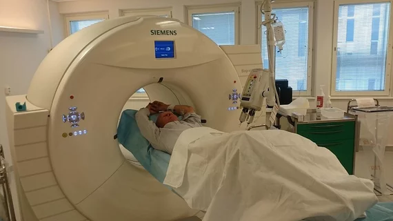
Molecular Imaging

Molecular imaging (also called nuclear medicine or nuclear imaging) can image the function of cells inside the body at the molecular level. This includes the imaging modalities of positron emission computed tomography (PET) and single photon emission computed tomography (SPECT) imaging. How does PET and SPECT imaging work? Small amounts of radioactive material (radiopharmaceuticals) injected into a patient. These can use sugars or chemical traits to bond to specific cells. The radioactive material is taken up by cells that consume the sugars. The radiation emitted from inside the body is detected by photon detectors outside the body. Computers take the data to assemble images of the radiation emissions. Nuclear images may appear fuzzy or ghostly rather than the sharper resolution from MRI and CT.  But, it provides metabolic information at a cellular level, showing if there are defects in the function of the heart, areas of very high metabolic activity associated with cancer cells, or areas of inflammation, data not available from other modalities. These noninvasive imaging exams are used to diagnose cancer, heart disease, Alzheimer’s and Parkinson’s disease, bone disorders and other disorders. 

Thumbnail

Optical metabolic imaging could be ideal for breast cancer research

Fluorescence optical imaging of the breast has the potential to capture dynamic metabolic response signaling malignancy, according to a review published online Oct. 15 in Cancer Research.

New funding for intraoperative optical breast imaging

Lightpoint Medical and Sagentia Technology and Product Development have been awarded a $200,000 grant by the U.K.’s Technology Strategy Board, the company announced earlier this month.

PET/CT: C-reactive protein could be a precursor to cardiovascular disease

FDG uptake rose substantially in PET/CT scans of patients with higher levels of C-reative protein in their carotid arteries, pinpointing a potential biomarker for cardiovascular disease, according to an article published online Oct. 31 in the Journal of Nuclear Medicine.

Y-90 microspheres extend survival in 50 percent of metastatic colorectal cancer patients

Salvage patients, those with colorectal metastases to the liver who do not to respond to both first and second lines of treatment, are responding significantly to radioembolization with Y-90 microspheres. The dual-action treatment has been shown to lead to more than 12 months of overall survival, according to a study published Nov. 1 in the Journal of Nuclear Medicine.

Thumbnail

Celebrating International Day of Radiology

It is International Day of Radiology Nov. 8 and radiological societies and institutions around the world are commemorating historical benchmarks and technological advancements in the field.

Fluorine-labeled bombesin PET agent a winner for prostate cancer imaging

In a comparison of gastrin-releasing peptide receptor (GRPR) bombesin analogs, F-18 aluminum flouride-labeled NODAGA-RM1 showed the most promise for PET imaging of prostate cancer, according to a study published online Nov. 6 in the Journal of Nuclear Medicine.

Being bilingual could delay onset of dementia

People fluent in two languages may be warding off the cognitive decline associated with dementia for up to six years longer than single-language speakers, according to a study published online Nov. 6 in Neurology.

Thumbnail

PQRS: Play Now or Pay Later

Human nature being what it is, physician participation in Medicare’s Physician Quality Reporting System (PQRS) will likely accelerate as the agency phases incentives out and penalties in. The effect may prove especially conspicuous since the bonuses have been voluntary and modest. By contrast, the forfeitures will be automatic and, if paired with other pay-for-performance requirements, impossible to ignore.