Molecular Imaging

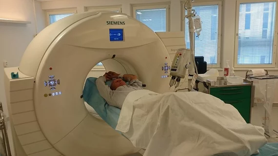
Molecular imaging (also called nuclear medicine or nuclear imaging) can image the function of cells inside the body at the molecular level. This includes the imaging modalities of positron emission computed tomography (PET) and single photon emission computed tomography (SPECT) imaging. How does PET and SPECT imaging work? Small amounts of radioactive material (radiopharmaceuticals) injected into a patient. These can use sugars or chemical traits to bond to specific cells. The radioactive material is taken up by cells that consume the sugars. The radiation emitted from inside the body is detected by photon detectors outside the body. Computers take the data to assemble images of the radiation emissions. Nuclear images may appear fuzzy or ghostly rather than the sharper resolution from MRI and CT.  But, it provides metabolic information at a cellular level, showing if there are defects in the function of the heart, areas of very high metabolic activity associated with cancer cells, or areas of inflammation, data not available from other modalities. These noninvasive imaging exams are used to diagnose cancer, heart disease, Alzheimer’s and Parkinson’s disease, bone disorders and other disorders. 

FDG PET hot spots spell risk of rupture in abdominal aortic aneurysm

A higher risk of aneurysm is on the menu for patients who test positive for abdominal aortic inflammation and other cellular changes, according to a study published online Sept. 5 in the Journal of Nuclear Medicine.

PET/CT provides snapshot of E. coli coronary stent infection

Infection imaging with PET/CT is gaining traction and could help detect serious complications after coronary stent placement, as evidenced by an E. coli case study published online Aug. 24 in the Journal of Nuclear Cardiology.

FDA clears Siemens’ Symbia Intevo integrated SPECT and CT system

Siemens Healthcare has announced that the FDA has granted 510(k) clearance for Symbia Intevo—the world’s first xSPECT system, which combines the high sensitivity of single-photon emission computed tomography (SPECT) with the high specificity of CT.

Metabolic PET imaging provides clues to developing Huntington’s disease

A functional neural network is under investigation as a major player in the progression of Huntington’s disease. Metabolic imaging of this region could serve as a tool for future treatments, according to a study published online Aug. 29 in the Journal of Clinical Investigation.

Chasing the lightning: Molecular epilepsy imaging with PET and SPECT

Recent studies have indicated that epilepsy may be more comprehensively characterized with the help of PET and SPECT, and these modalities could benefit the 1-2 percent of the U.S. population with the neurological disorder, according to a review published online Aug 22 in the Journal of Nuclear Medicine.

Thumbnail

Estrogen receptor PET imaging detects early stage breast cancer, with limitations

Early-stage breast cancer lesions can be detected with F-18 FES PET, but success depends on tumor size and level of disease, according to a study published online Aug. 22 in the Journal of Nuclear Medicine.

PET on a Leash: Reimbursement Developments and Challenges

The operative phrase for PET reimbursement circa 2013 may be “cautiously optimistic.”

Radiation Exposure: Connecting the Dots

The right dose to the right patient at the right time is the mantra. But tracking dose is gaining in popularity among physicians and patients.