Molecular Imaging

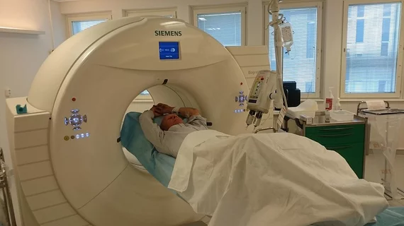
Molecular imaging (also called nuclear medicine or nuclear imaging) can image the function of cells inside the body at the molecular level. This includes the imaging modalities of positron emission computed tomography (PET) and single photon emission computed tomography (SPECT) imaging. How does PET and SPECT imaging work? Small amounts of radioactive material (radiopharmaceuticals) injected into a patient. These can use sugars or chemical traits to bond to specific cells. The radioactive material is taken up by cells that consume the sugars. The radiation emitted from inside the body is detected by photon detectors outside the body. Computers take the data to assemble images of the radiation emissions. Nuclear images may appear fuzzy or ghostly rather than the sharper resolution from MRI and CT.  But, it provides metabolic information at a cellular level, showing if there are defects in the function of the heart, areas of very high metabolic activity associated with cancer cells, or areas of inflammation, data not available from other modalities. These noninvasive imaging exams are used to diagnose cancer, heart disease, Alzheimer’s and Parkinson’s disease, bone disorders and other disorders. 

PhRMA reports 444 new medications in line for neurological diseases

Just under 450 new therapies are in the works for a wide range of neurological disorders, including Alzheimer’s  and Parkinson’s disease, brain cancer, epilepsy, multiple sclerosis and many others, according to a 2013 report released Wednesday by PhRMA, a biopharmaceutical industry trade group.

Protocol for the pipeline: Endpoints for PET tracer trials

Following two conferences held by the Medical Imaging & Technology Alliance (MITA) and a variety of stakeholders, a new focus on diagnostic PET tracers’ effect on patient management rather than direct health benefits is being presented for potential clinical trials, according to a review published August 1 in the Journal of Nuclear Medicine.

Hypoxia-abled biomarkers could be a boon for prostate cancer imaging

The gastrin-releasing peptide receptor (BB2r) has become a popular target in prostate cancer imaging, but low local retention of tracers at the tumor site impedes imaging. Hypoxia-enhanced targeting of these receptors may be just what the doctor ordered for better tumor-tracer retention, according to a study published July 29th in the Journal of Nuclear Medicine.

FDGPET found wanting in macrophage imaging of atherosclerosis

Novel molecular imaging detection of atherosclerosis is increasingly being presented to head off arterial damage and risk of cardiac events before they are found by more conventional methods, but FDGPET may not be the ideal solution, according to a study published July 25th in the Journal of Nuclear Medicine.

F-18 FDM may offer new oncologic PET agent

A fluorine-based PET agent on the workbench called 2-Deoxy-2-18F-fluoro-D-mannose (F-18 FDM) has been found to work similarly and potentially better than FDG for cancer imaging, especially of the brain, according to a study published July 10 in the Journal of Nuclear Medicine.

FDG PET/CT reveals necrotic nuances in sarcomas that predict survival

Little improvement in survival has been made for patients with sarcomas, despite an array of aggressive treatments. Researchers are trying to identify patients at high risk of recurrence by performing FDG PET/CT to quantify necrotic areas of tissue, a strong indicator of survival, according to research published in the July issue of the American Journal of Roentgenology.

Thumbnail

MR + PET highlight elusive triple negative breast cancers

The heterogeneity of breast cancer can make it difficult to capture, especially in cases where telltale receptor expression or tumor characterization are not involved. However, MR and F-18 FDG PET may be able to suss out the more obscure phenotypes of the disease, according to a study published July 17 in Radiology.

Software provides a library of radiopharmaceutical drug interactions

A newly developed software called Datinrad has combed through extensive literature and compiled more than two hundred radiopharmaceutical interactions and adverse effects is now accessible through a single PC portal, according to an article published July 15 in the Journal of Nuclear Medicine Technology.