Molecular Imaging

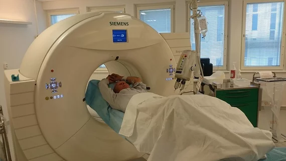
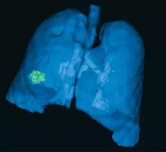
Molecular imaging (also called nuclear medicine or nuclear imaging) can image the function of cells inside the body at the molecular level. This includes the imaging modalities of positron emission computed tomography (PET) and single photon emission computed tomography (SPECT) imaging. How does PET and SPECT imaging work? Small amounts of radioactive material (radiopharmaceuticals) injected into a patient. These can use sugars or chemical traits to bond to specific cells. The radioactive material is taken up by cells that consume the sugars. The radiation emitted from inside the body is detected by photon detectors outside the body. Computers take the data to assemble images of the radiation emissions. Nuclear images may appear fuzzy or ghostly rather than the sharper resolution from MRI and CT.  But, it provides metabolic information at a cellular level, showing if there are defects in the function of the heart, areas of very high metabolic activity associated with cancer cells, or areas of inflammation, data not available from other modalities. These noninvasive imaging exams are used to diagnose cancer, heart disease, Alzheimer’s and Parkinson’s disease, bone disorders and other disorders. 

Radiation Exposure: Connecting the Dots

The right dose to the right patient at the right time is the mantra. But tracking dose is gaining in popularity among physicians and patients.

PEM: An Additional Arrow In the Breast Imaging Quiver

Many physicians and patients cast staging breast MRI as an imperfect exam. From the physician’s perspective, the exam generates thousands of difficult-to-interpret images. False-positive findings are not uncommon.

Cancer Care Informatics: Challenges In Nuclear Medicine

Multiple radiopharmaceuticals in the development pipeline are showing real promise in the realms of oncology, neurology and cardiology. 

The Proliferation of Activity and Meetings in MI

This spring and summer was a very busy one in the field of molecular imaging. Starting in May, we had the International Society of Radiopharmaceutical Sciences meeting in Jeju, South Korea. 

MaineHealth aims to achieve American Society of Nuclear Cardiology (ASNC) guidelines with UltraSPECT technology

Healthcare system selects UltraSPECT nuclear medicine software for lowering dose and reducing exam time.

‘Betabox’ micro molecular imaging system could spur drug trials

In vitro drug trials could get a boost from a micrometer-small molecular imaging system that captures cells and tests their response to a miniscule flood of radiopharmaceuticals, according to a study published Aug. 26 in the Journal of Nuclear Medicine.

Molecular imaging industry set for slow-going growth

At least 14 new molecular imaging agents are being groomed for clinical use, with approval of three major radiotracers expected by 2016, according to an Aug. 2013 GlobalData report entitled “The Future of Molecular Imaging.”

FDG PET/CT predicts bone cancer patient survival

FDG PET/CT imaging prior to osteosarcoma surgery and chemotherapy may be able to provide a clearer picture of patient risk and prognosis, according to a study published Aug. 15 in the Journal of Nuclear Medicine.