Molecular Imaging

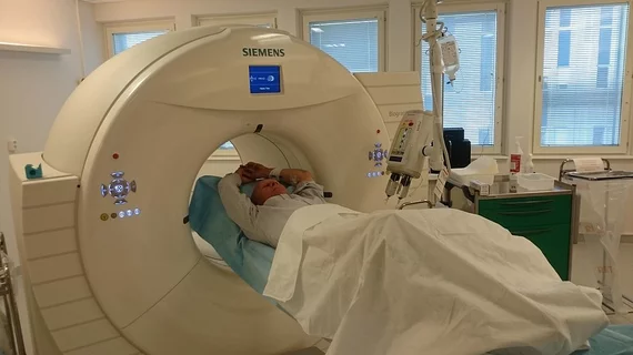
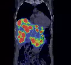
Molecular imaging (also called nuclear medicine or nuclear imaging) can image the function of cells inside the body at the molecular level. This includes the imaging modalities of positron emission computed tomography (PET) and single photon emission computed tomography (SPECT) imaging. How does PET and SPECT imaging work? Small amounts of radioactive material (radiopharmaceuticals) injected into a patient. These can use sugars or chemical traits to bond to specific cells. The radioactive material is taken up by cells that consume the sugars. The radiation emitted from inside the body is detected by photon detectors outside the body. Computers take the data to assemble images of the radiation emissions. Nuclear images may appear fuzzy or ghostly rather than the sharper resolution from MRI and CT.  But, it provides metabolic information at a cellular level, showing if there are defects in the function of the heart, areas of very high metabolic activity associated with cancer cells, or areas of inflammation, data not available from other modalities. These noninvasive imaging exams are used to diagnose cancer, heart disease, Alzheimer’s and Parkinson’s disease, bone disorders and other disorders. 

FDA ushers in Sorafenib for the treatment of metastatic thyroid cancer

Sorafenib, otherwise known as Nevaxar, is now approved to treat advanced thyroid cancer, as of a Nov. 22 statement from the FDA.

South African plant leak has caused worldwide medical isotope shortage

South African Nuclear Energy Corp. announced this month that a leak has resulted in a global shortage of molybdenum-99, the precursor for Tc-99m used in a majority of molecular imaging scans. Authorities reportedly conducted an investigation Nov. 12 at the NTP plant in Pelindaba, just shy of 20 miles from the capital of Pretoria. It was estimated that the leak began Nov. 2. and was comprised of noble gas and iodine. Repairs are underway and the NTP plant will await independent review and authorization from the National Nuclear Regulator before operation can begin again.

FDA gives orphan drug designation for Ga-68 DOTATOC

Gallium-68 DOTATOC is now blazing faster trail toward U.S. regulatory approval for neuroendocrine cancer imaging, according to a Nov. 18 statement from the Society of Nuclear Medicine and Molecular Imaging (SNMMI). 

Four times longer half-life with Sc-44 DOTA oncologic PET agent

A longer lasting alternative to gadolinium-68 for PET cancer imaging prior to radionuclide therapy has been found in a cyclotron-produced scandium-44 agent, according to a study published online Nov. 6 in the Journal of Nuclear Medicine.

Quantum dots facilitate a leap into alternative energy

The semiconductor photocatalysts known in molecular imaging as "quantum dots" are now being used for something completely different: alternative energy. The nanocrystals, instead of highlighting tissues, are harnessing methanol through a process of photosynthesis. This and other research, including advances in biomedical 3D printing and oncologic iron-based MRI contrast agents are featured in a profile of new research published in the New York Academy of Sciences magazine.

Thumbnail

Optical metabolic imaging could be ideal for breast cancer research

Fluorescence optical imaging of the breast has the potential to capture dynamic metabolic response signaling malignancy, according to a review published online Oct. 15 in Cancer Research.

New funding for intraoperative optical breast imaging

Lightpoint Medical and Sagentia Technology and Product Development have been awarded a $200,000 grant by the U.K.’s Technology Strategy Board, the company announced earlier this month.

PET/CT: C-reactive protein could be a precursor to cardiovascular disease

FDG uptake rose substantially in PET/CT scans of patients with higher levels of C-reative protein in their carotid arteries, pinpointing a potential biomarker for cardiovascular disease, according to an article published online Oct. 31 in the Journal of Nuclear Medicine.