Molecular Imaging

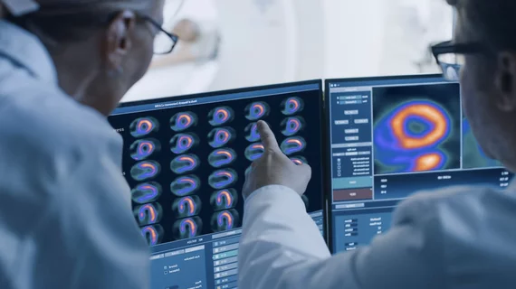
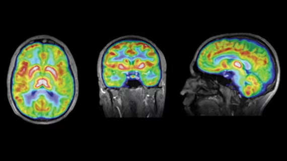
Molecular imaging (also called nuclear medicine or nuclear imaging) can image the function of cells inside the body at the molecular level. This includes the imaging modalities of positron emission computed tomography (PET) and single photon emission computed tomography (SPECT) imaging. How does PET and SPECT imaging work? Small amounts of radioactive material (radiopharmaceuticals) injected into a patient. These can use sugars or chemical traits to bond to specific cells. The radioactive material is taken up by cells that consume the sugars. The radiation emitted from inside the body is detected by photon detectors outside the body. Computers take the data to assemble images of the radiation emissions. Nuclear images may appear fuzzy or ghostly rather than the sharper resolution from MRI and CT.  But, it provides metabolic information at a cellular level, showing if there are defects in the function of the heart, areas of very high metabolic activity associated with cancer cells, or areas of inflammation, data not available from other modalities. These noninvasive imaging exams are used to diagnose cancer, heart disease, Alzheimer’s and Parkinson’s disease, bone disorders and other disorders. 

'3D nanoSIMS' molecular imaging system touts nano resolutions

A new label-free molecular imaging system called the 3D nanoSIMS could provide superior drug research studies at intensely tiny spatial resolutions, announced the National Physical Laboratory in Middlesex, United Kingdom, on Dec. 10.

Benzamide imaging agent could lead to a novel therapy for metastatic melanoma

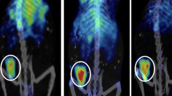
Average survival for metastatic melanoma patients is less than 5 percent after five years—a disheartening figure, but an investigational agent incorporating benzamide in a melanin-targeted SPECT radiotracer could change the numbers, according to a study published Nov. 16 in the Journal of Nuclear Medicine.

Take heart: PET market to increase with new cardiac agents

Two novel cardiovascular PET agents are in the wings from FluoroPharma Medical, according to a recent profile in Forbes magazine.

Thumbnail

Clusterin and amyloid may be linked in Alzheimer’s brain atrophy

The usual subjects in the Alzheimer’s brain are patterns of beta-amyloid plaque and tau aggregation, but a third perpetrator, clusterin, may be playing a more important role in the development of the disease than previously thought, according to a study published Dec. 30 in JAMA Neurology.

Ketamine boosts serotonin in PET study

The anesthetic ketamine, used in both humans and animals, may have a new use: treating chronic, drug resistant major depression, according to a study published Jan. 7 in Translational Psychiatry.

Isn’t it sweet: FDG may have a rival

The name is fluorodeoxymannose (FDM) and it is giving FDG some competition in the realm of glucose analogue imaging of atherosclerosis, according to a report published Jan. 12 in Nature.

Thumbnail

Coming of age for gallium-68 neuroendocrine imaging

Long past are the days when fluorodeoxyglucose was the primary workhorse for imaging neuroendocrine tumors (NETs). Now there is a veritable cornucopia of agents to choose from, but a family of compounds radiolabeled with gallium-68 have been gaining a lot of headway. These are Ga-68 DOTANOC, DOTATOC and DOTATATE.

‘Blinking’ DNA probes revolutionize optical microscopy

Researchers are developing probes that highlight the goings on inside cells at a super high resolution at Wyss Institute for Biologically Inspired Engineering of Harvard University in Boston.