Molecular Imaging

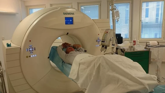
Molecular imaging (also called nuclear medicine or nuclear imaging) can image the function of cells inside the body at the molecular level. This includes the imaging modalities of positron emission computed tomography (PET) and single photon emission computed tomography (SPECT) imaging. How does PET and SPECT imaging work? Small amounts of radioactive material (radiopharmaceuticals) injected into a patient. These can use sugars or chemical traits to bond to specific cells. The radioactive material is taken up by cells that consume the sugars. The radiation emitted from inside the body is detected by photon detectors outside the body. Computers take the data to assemble images of the radiation emissions. Nuclear images may appear fuzzy or ghostly rather than the sharper resolution from MRI and CT.  But, it provides metabolic information at a cellular level, showing if there are defects in the function of the heart, areas of very high metabolic activity associated with cancer cells, or areas of inflammation, data not available from other modalities. These noninvasive imaging exams are used to diagnose cancer, heart disease, Alzheimer’s and Parkinson’s disease, bone disorders and other disorders. 

Medical sensor market expected to reach $15.5B in five years

The industry for medical sensors, which includes diagnostic imaging and disease monitoring, was valued at $10.1 billion in 2012 and is expected to grow significantly by 2019, according to an industry sector report published Dec. 5 by Transparency Market Research.

Thumbnail

Value of amyloid imaging is ‘uncertain’

A sweep of more than 550 articles from the medical literature on Alzheimer’s disease diagnosis left reviewers questioning the utility of a positive beta-amyloid scan, according to a meta-analysis published in the January issue of JAMA Internal Medicine. 

Discerning infection from inflammation: A new PET/CT technique

Antimicrobial peptide PET/CT imaging with gallium-68 appears to break through previous roadblocks in differentiating infections and sterile inflammatory processes, according to a study published Jan. 16 in the Journal of Nuclear Medicine.

Thumbnail

Lab on the field: Combining molecular imaging and microfluidics

A tiny molecular imaging system could be used on the field to measure brain injury or in the lab to capture individual molecules that betray the biology of cancer, neurodegenerative diseases such as Alzheimer's and viruses like human immunodeficiency virus (HIV). 

Thumbnail

Playing fluorescent ‘tag’ to study the biology of memory

Never-before-seen preclinical fluorescence imaging has broken new ground in the understanding of how memories are formed by nerve cells in the brain.

Alzheimer’s partnership to fund new research in the United Kingdom

A partnership between the United Kingdom-based Alzheimer’s Society and the Alzheimer’s Drug Discovery Foundation is engaged to fund up to $1.5 million for Alzheimer’s drug development programs.

Thumbnail

Biomedical imaging goes subatomic with advanced electron microscopy

Electron microscopy researchers have been given the Banco Bilbao Vizcaya Argentaria (BBVA) Foundation Frontiers of Knowledge Award for pushing the technology into the picometer range—or one-hundredth the diameter of a hydrogen atom.

SMARTVis improves diagnostics by fusing cardiac CTA and SPECT MPI data

Combining CT angiography (CTA) and SPECT MPI is useful for the evaluation of coronary artery disease (CAD) and shows more diagnostic benefit than just one of the modalities or consecutive use of the two technologies, according to a study published Jan. 1 in the Journal of Nuclear Medicine.