Molecular Imaging

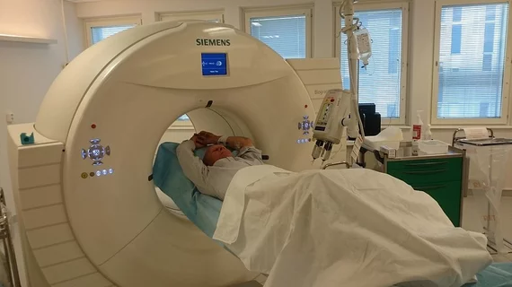
Molecular imaging (also called nuclear medicine or nuclear imaging) can image the function of cells inside the body at the molecular level. This includes the imaging modalities of positron emission computed tomography (PET) and single photon emission computed tomography (SPECT) imaging. How does PET and SPECT imaging work? Small amounts of radioactive material (radiopharmaceuticals) injected into a patient. These can use sugars or chemical traits to bond to specific cells. The radioactive material is taken up by cells that consume the sugars. The radiation emitted from inside the body is detected by photon detectors outside the body. Computers take the data to assemble images of the radiation emissions. Nuclear images may appear fuzzy or ghostly rather than the sharper resolution from MRI and CT.  But, it provides metabolic information at a cellular level, showing if there are defects in the function of the heart, areas of very high metabolic activity associated with cancer cells, or areas of inflammation, data not available from other modalities. These noninvasive imaging exams are used to diagnose cancer, heart disease, Alzheimer’s and Parkinson’s disease, bone disorders and other disorders. 

Thumbnail

Brilliant Signs: Advancements in Brain & Neuroendocrine Imaging

The past year has been a groundbreaking one for neurology. 

The Logical Next Step

When I began my PhD research almost 40 years ago, nuclear medicine research was almost entirely focused on diagnosis. 

AAA acquires 100% share of UK nuclear medicine company

Advanced Accelerator Applications (AAA), a major molecular medicine company based in Bourg en Bresse, France, announced Mar. 4 that the firm had acquired 100 percent of shares of the privately held U.K.-based Imaging Equipment, a distributor of nuclear medicine technologies and radiopharmaceuticals across the U.K. and Ireland.

Popular media may be kicking up inappropriate PET use

The question of appropriate use of advanced nuclear medicine procedures is more salient than it has ever been, with overall health cost under government scrutiny and imaging eyed in particular due to increasing procedure numbers. Some experts are pointing to non-medical news sites and other media as a culprit in patient demand for potentially inappropriate PET use, according to a study published Feb. 18 in Cancer Epidemiology, Biomarkers & Prevention.

PET/MR: still struggling to solidify clinical practice

There are two main questions that remain on the minds of those evaluating PET/MR for clinical practice: Is PET/CT just as good? If yes, isn’t PET/CT cheaper? These questions are answered with a resounding ‘sometimes,’ according to a review published Feb. 20 in the Journal of Nuclear Medicine.

Biomolecule optical imaging trumps fluorescence for the small

Imaging small biomolecules in living cells and animals just took a huge leap over the conventional restrictions of fluorescence imaging. Scientists at Columbia University in New York City have developed a means of observing miniscule molecular materials using novel chemistry and physics.

Thumbnail

Alzheimer’s research: Culprit of deadly tau aggregation found

Understanding tau behavior in Alzheimer’s disease could alter the course of diagnostics and therapy development. Research conducted in part by scientists at the Technische Universitat Munchen (TUM) has revealed an essential mechanism of tau deposition and neurodegenerative disease, the institution announced today.

Aptamer-based molecular agents have arrived

Biomarkers built upon aptamers rather than peptides and antibodies offer multiple advantages for molecular imaging and several are currently under development, according to a review published Feb. 13 in the Journal of Nuclear Medicine.