Molecular Imaging

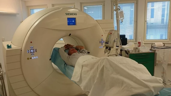
Molecular imaging (also called nuclear medicine or nuclear imaging) can image the function of cells inside the body at the molecular level. This includes the imaging modalities of positron emission computed tomography (PET) and single photon emission computed tomography (SPECT) imaging. How does PET and SPECT imaging work? Small amounts of radioactive material (radiopharmaceuticals) injected into a patient. These can use sugars or chemical traits to bond to specific cells. The radioactive material is taken up by cells that consume the sugars. The radiation emitted from inside the body is detected by photon detectors outside the body. Computers take the data to assemble images of the radiation emissions. Nuclear images may appear fuzzy or ghostly rather than the sharper resolution from MRI and CT.  But, it provides metabolic information at a cellular level, showing if there are defects in the function of the heart, areas of very high metabolic activity associated with cancer cells, or areas of inflammation, data not available from other modalities. These noninvasive imaging exams are used to diagnose cancer, heart disease, Alzheimer’s and Parkinson’s disease, bone disorders and other disorders. 

ECR 2014: EuroSafe Imaging campaign launched by ESR

As the European Congress of Radiology (ECR) 2014 kicked off yesterday, the founding European Society of Radiology (ESR) launched a partnership and project with the American College of Radiology, officially titled EuroSafe Imaging, with an aim to improve adoption of clinical protocols for safer, more appropriate use of radiological procedures.

Thumbnail

ECR 2014: Future of molecular imaging is now

VIENNA—Today marked the opening of the European Congress of Radiology (ECR) 2014 being held March 6-10 at the Austria Center Vienna. This is the 20th annual meeting of the European Society of Radiology (ESR), where an estimated 20,000 participants from 100 countries have come together to discuss the latest news and developments in the worldwide radiological community.

Thumbnail

On the Swell: Imaging Infection & Inflammation

Inflammation plays a major role in disease processes.

Thumbnail

Dialing Down Dose: Radiation Tracking and Reporting Tools

As imaging studios have markedly increased in the past two decades, so has the concern among some clinicians and media surrounding radiation dose management.

Thumbnail

Revolution, Not Revolt: The New Era of Stem Cell Cancer Research

Irving L. Weissman, MD’s stem cell research using PET and optical imaging has revealed how a novel antibody-based biomarker highlights areas of residual cancer with broad potential for therapeutic use.

Thumbnail

SPECT & PET: Tailoring Heart Failure Therapy

Molecular imaging is beginning to give cardiologists insight into biological processes behind heart failure, knowledge that may allow them to monitor disease progression and tailor therapies. But market realities still pose barriers.

Thumbnail

Alzheimer’s Imaging Agents: The Regulatory Horizon

“Alzheimer’s disease is probably the most underfunded disease in the U.S.,” charges Dean M. Hartley, PhD, director of science initiatives, medical and scientific relations, for the Alzheimer’s Association.

Thumbnail

High-tech Modeling: Computer Mockups Hone in on Human Physiology

An exploration of recent projects at Argonne National Laboratory and Johns Hopkins University traverse the numerical and graphical landscape of cerebral blood flow, the pathology of malaria, brain aneurysm, sickle cell anemia and even cancer.