Molecular Imaging

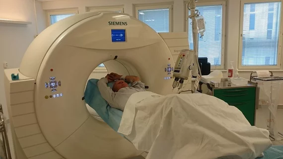
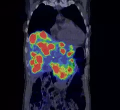
Molecular imaging (also called nuclear medicine or nuclear imaging) can image the function of cells inside the body at the molecular level. This includes the imaging modalities of positron emission computed tomography (PET) and single photon emission computed tomography (SPECT) imaging. How does PET and SPECT imaging work? Small amounts of radioactive material (radiopharmaceuticals) injected into a patient. These can use sugars or chemical traits to bond to specific cells. The radioactive material is taken up by cells that consume the sugars. The radiation emitted from inside the body is detected by photon detectors outside the body. Computers take the data to assemble images of the radiation emissions. Nuclear images may appear fuzzy or ghostly rather than the sharper resolution from MRI and CT.  But, it provides metabolic information at a cellular level, showing if there are defects in the function of the heart, areas of very high metabolic activity associated with cancer cells, or areas of inflammation, data not available from other modalities. These noninvasive imaging exams are used to diagnose cancer, heart disease, Alzheimer’s and Parkinson’s disease, bone disorders and other disorders. 

Johnson and Johnson bankrolling Alector Alzheimer’s therapy

The Johnson and Johnson Innovation Center is bankrolling a research project with San Francisco-based Alector to prove feasibility of a monoclonal antibody agent that homes in on and treats Alzheimer’s disease processes in the brain, Alector announced yesterday.

Thumbnail

Almost $3M granted for Lightpoint intraoperative imaging clinical trials

A grant amounting to $2.8 million has been awarded to Lightpoint Medical and surgical partners of the company headquartered in Rickmansworth, England, to bolster clinical trials for intraoperative imaging systems for prostate and breast cancer.

FLT PET and MR spectroscopy: The voxels do the talking for high-grade gliomas

A voxel-based comparison of F-18 fluorothymidine (FLT) PET and MR spectroscopy revealed good contrast between tumorous and healthy tissues for both methods, according to a study presented during the scientific session of the 2014 European Congress of Radiology (ECR), which concludes today at Austria Center Vienna.

FET PET points to additional glioma activity compared with MRI

PET provides more detail than perfusion-weighted MRI about glioma tumor extent and metabolic activity, according to a study published online Feb. 27 in the Journal of Nuclear Medicine.

Smoking while pregnant diminishes baby’s brain size, gray matter

Prenatal tobacco use adversely affects brain development by limiting a child’s total brain volume and particularly their gray matter, according to longitudinal MRI research published in the March 2014 edition of Neuropsychopharmacology.

Thumbnail

ECR 2014: PEM shows both high specificity and sensitivity for breast cancer

VIENNA—Positron emission mammography (PEM) was found to have 100 sensitivity for all imaged breast tumors, according to a study being presented during the 2014 European Congress of Radiology (ECR) scientific sessions.

ECR 2014: Diffusion tensor imaging predicts motor function recovery following stroke

VIENNA—Stroke victims often lose significant motor function as a result of acute ischemic attacks. Diffusion tensor imaging (DTI) has the potential to predict just how much of that motor function patients will likely recover over time, according to a scientific presentation at the 2014 European Congress of Radiology (ECR).

ECR 2014: First-in-human glutamine PET trial results revealed

Preliminary data regarding the first known human trial involving glutamine PET for the detection of glioma showed encouraging pharmacokinetics and biodistribution, according to researchers presenting at this year’s European Congress of Radiology (ECR).