Molecular Imaging

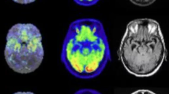
Molecular imaging (also called nuclear medicine or nuclear imaging) can image the function of cells inside the body at the molecular level. This includes the imaging modalities of positron emission computed tomography (PET) and single photon emission computed tomography (SPECT) imaging. How does PET and SPECT imaging work? Small amounts of radioactive material (radiopharmaceuticals) injected into a patient. These can use sugars or chemical traits to bond to specific cells. The radioactive material is taken up by cells that consume the sugars. The radiation emitted from inside the body is detected by photon detectors outside the body. Computers take the data to assemble images of the radiation emissions. Nuclear images may appear fuzzy or ghostly rather than the sharper resolution from MRI and CT.  But, it provides metabolic information at a cellular level, showing if there are defects in the function of the heart, areas of very high metabolic activity associated with cancer cells, or areas of inflammation, data not available from other modalities. These noninvasive imaging exams are used to diagnose cancer, heart disease, Alzheimer’s and Parkinson’s disease, bone disorders and other disorders. 

Thumbnail

Upcoming ECR 2014 in Vienna ramps up radiogenomics

The European Congress of Radiology (ECR) in Vienna, the largest radiological conference throughout Europe, will be opening in less than two weeks from March 6-10 at the Austria Center Vienna, where it has now been held for 20 consecutive years.

ADA Medical moves into preclinical research

The Toronto-based research group ADA Medical announced Feb. 14 that it would be collaborating with Ephoran Multi-Imaging Solutions in Turin, Italy, to provide preclinical imaging services.

Cancer disparity gets a closer look

Major cancer research organizations are banding together to assess how cancer disparity research will move forward. In the first collaboration of its kind, the American Association for Cancer Research (AACR) announced Feb. 13 that the institute would be working with the American Cancer Society, the American Society of Clinical Oncology (ASCO) and the National Cancer Institute (NCI) to address concerns such as access to health care and factors related to variability of cancer outcomes.

Thumbnail

RNA-enabled nanoparticles could treat liver disease

Massachusetts Institute of Technology engineers have fashioned a smarter nanoparticle that runs RNA interference as a means of disease modification, the institute announced Feb. 10.

Microfluidics could map how nanoparticles interact with atherosclerosis

The use of an endothelialized microfluidic chip could be the key to understanding how nanoparticles in biomedicine behave in the presence of atherosclerosis, according to a study published online Jan. 6 in the Proceedings of the National Academy of Sciences.

Thumbnail

Precious Cargo: Nanotechnology enlists silk, diamonds and gold to improve molecular medicine

Nanomedicine incorporates some of the most prized materials on earth in order to improve not only its biocompatibility and imaging potential, but its ability to sneak up on and change the way that cells function for the tiniest aspect of personalized medicine.

New $15M biomarker lab planned for Singapore

Los Angeles-based ImaginAb is slated to collaborate with Duke-NUS Graduate School of Medicine and National Research Foundation Corp Lab on a $15 million imaging biomarker lab, ImaginAb announced Feb. 10.

Sedecal and Xstrahl partner on pre-clinical systems

A distribution contract has been struck between preclinical molecular imaging system manufacturer Sedecal headquartered in Spain and United Kingdom radiation oncology systems manufacturer Xstrahl Life Sciences, the company announced today.