Molecular Imaging

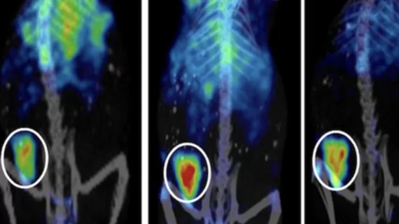
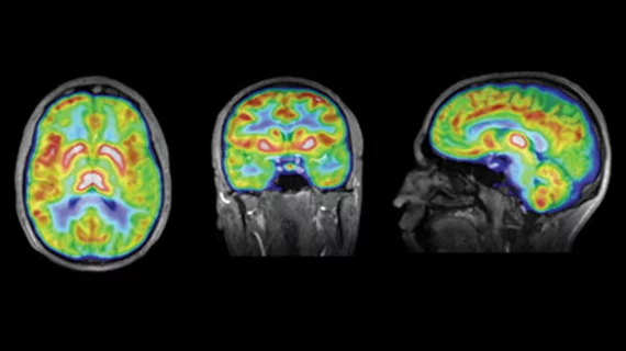
Molecular imaging (also called nuclear medicine or nuclear imaging) can image the function of cells inside the body at the molecular level. This includes the imaging modalities of positron emission computed tomography (PET) and single photon emission computed tomography (SPECT) imaging. How does PET and SPECT imaging work? Small amounts of radioactive material (radiopharmaceuticals) injected into a patient. These can use sugars or chemical traits to bond to specific cells. The radioactive material is taken up by cells that consume the sugars. The radiation emitted from inside the body is detected by photon detectors outside the body. Computers take the data to assemble images of the radiation emissions. Nuclear images may appear fuzzy or ghostly rather than the sharper resolution from MRI and CT.  But, it provides metabolic information at a cellular level, showing if there are defects in the function of the heart, areas of very high metabolic activity associated with cancer cells, or areas of inflammation, data not available from other modalities. These noninvasive imaging exams are used to diagnose cancer, heart disease, Alzheimer’s and Parkinson’s disease, bone disorders and other disorders. 

Novel immuno-PET agent seeks cancer vasculature

An investigational immune-PET biomarker maps tumor vasculature while skipping healthy tissues, according to a study published Feb. 13 in the Journal of Nuclear Medicine.

Dual therapy proven to improve survival in patients with glioma

Following radiation therapy with chemotherapy has now been shown to prolong survival for glioma patients. Results of the study were announced Feb. 3 by the National Institutes of Health.

Edinburgh Molecular Imaging gets more than $6M in funding for optical innovation

The Scottish developer of optical molecular imaging technology, Edinburgh Molecular Imaging, has received venture capital funding amounting to more than $6 million, the University of Edinburgh announced Feb 7.

Thumbnail

Improving breast cancer outcomes: FDG PET peers in on adrenoceptor blockers

Adrenoceptor blockers have been found to stave off metastasis and recurrence of breast cancer. This may be due to their interruption of key genes involved in glucose metabolism as visualized by FDG-PET, according to a study published Feb. 6 in the Journal of Nuclear Medicine.

Thumbnail

PET/MR is a toss-up for thoracic staging of non-small cell lung cancer

PET/MR doesn’t appear to provide any additional insight beyond PET/CT for thoracic staging of non-small cell lung cancer, according to a study published Feb. 6 in the Journal of Nuclear Medicine.

Advocacy trifecta funds neurodegenerative disease research

Three major patient advocacy associations, the Alzheimer’s Association, the Michael J. Fox Foundation and the W. Garfield Weston Foundation, have joined forces to encourage researchers to explore the biological bridges between Alzheimer’s, Parkinson’s and other neurodegenerative diseases.

Worldwide medical imaging reagents market on the rise

The global market for medical reagents, including those used for molecular imaging purposes, is expected to grow substantially through 2016, according to a market report announced Feb. 5 from research firm TechNavio.

Preliminary results look positive for cardiac fatty acid PET imaging

FluoroPharma Medical, based in Montclair, N.J., announced yesterday that new results from an ongoing phase II clinical trial for F-18 FCPHA cardiac PET for the diagnosis of acute coronary artery disease (CAD) were positive and providing validation for further research.