Magnetic Resonance Imaging

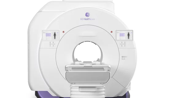
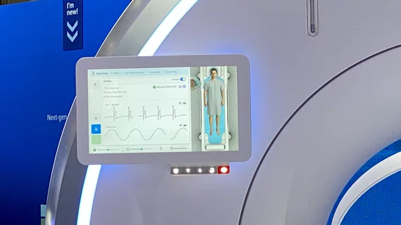
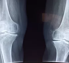
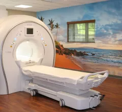
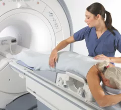
Magnetic resonance imaging (MRI) is the gold standard imaging modality for soft tissues. It produces detail cross-sectional images of soft tissue and bone anatomy, including muscles, tendons, ligaments, brain and organs, without the use of ionizing radiation. In addition to orthopedic imaging, MRI is also used for heart, brain and breast. MRI uses gadolinium contrast in many exams to highlight tissues and blood vessels, which enhances images and offers better diagnostic quality. It can also be used in conjunction with PET scans. How does MRI work? MR creates images by using powerful magnets to polarize hydrogen atoms in water (the body is made of of more than 80% water) so they face in one direction. A radiofrequency pulse is then used to ping these atoms, causing them to wobble, or resonate. The MRI coils detect this and computers can assemble images from the signals. Basic MRI scans will focus on the resonance of fat and water in two different sequences, which highlight and contrast different features in the anatomy.

Thumbnail

MRI an effective first step for imaging young patients with suspected acute appendicitis

MRI provides value as an initial imaging technique for pediatric patients with suspected acute appendicitis, according to a new study published in Radiology.

Thumbnail

Providers need consensus guidelines for follow-up imaging after benign MRI-guided breast biopsies

More consistent follow-up protocols after benign concordant MRI-guided percutaneous core needle biopsies (MR-PCNBs) could lead to better overall patient care, according to a case study published in the Journal of the American College of Radiology.

Thumbnail

A Brooklyn EMR Grows Toward Its Full Potential

Sponsored by FUJIFILM Healthcare Americas

Like every American academic healthcare institution, SUNY Downstate Medical Center in Brooklyn, N.Y., is a beehive of activity in three overlapping yet distinct areas of focus—patient care, physician education and medical research. 

Thumbnail

McGill University receives Canada's first whole-body 7T MRI scanner

The Montreal Neurological Institute and Hospital—also known as The Neuro—of McGill University in Montreal, Québec, Canada, has installed the country’s first whole-body 7-Tesla MRI scanner.

Thumbnail

How to improve prostate cancer detection during MRI-targeted transrectal US biopsies

Researchers have determined that increasing the number of core biopsy samples obtained using MRI-targeted transrectal ultrasound biopsy can lead to the detection of more clinically significant prostate cancers.

3T MRI scanner from Siemens Healthineers gains FDA clearance

Siemens Healthineers announced Wednesday, Jan. 30, that its MAGNETOM Lumina 3T MRI scanner has received FDA clearance.

Thumbnail

Hospital employee arrested after filming female patient undress for MRI

A ward boy working at a hospital in Pune, India—roughly three hours southwest of Mumbai—was arrested and charged for filming a female patient changing her clothes for an MRI scan, according to a report published online Jan. 27 by The Times of India. 

MRI suite in Illinois focuses on patient comfort

OSF Centers for Health in Peoria, Illinois, offers a wide-bore MRI that is four inches larger than a conventional scanner and includes a “Caring Suite” that makes it popular among patients.