Magnetic Resonance Imaging

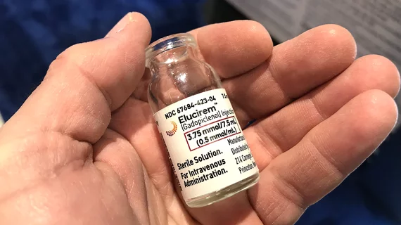
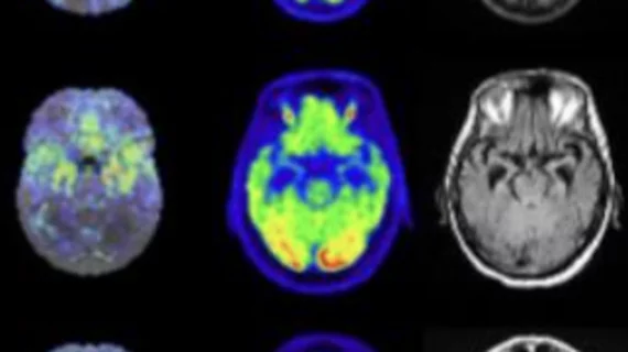
Magnetic resonance imaging (MRI) is the gold standard imaging modality for soft tissues. It produces detail cross-sectional images of soft tissue and bone anatomy, including muscles, tendons, ligaments, brain and organs, without the use of ionizing radiation. In addition to orthopedic imaging, MRI is also used for heart, brain and breast. MRI uses gadolinium contrast in many exams to highlight tissues and blood vessels, which enhances images and offers better diagnostic quality. It can also be used in conjunction with PET scans. How does MRI work? MR creates images by using powerful magnets to polarize hydrogen atoms in water (the body is made of of more than 80% water) so they face in one direction. A radiofrequency pulse is then used to ping these atoms, causing them to wobble, or resonate. The MRI coils detect this and computers can assemble images from the signals. Basic MRI scans will focus on the resonance of fat and water in two different sequences, which highlight and contrast different features in the anatomy.

Thumbnail

MRI scans help researchers understand the biology of autism

With the help of MRI, San Diego State University researchers found the amygdala of children on the autism spectrum disorder (ASD) had weaker connections with some regions of the brain compared to “typically developing” children within the same age group.

MRI shows potential to correct slow gastric emptying, gastroparesis

Researchers have used MRI to show the impact of sending electrical pulses to the vagus nerve in an attempt to correct gastrointestinal problems and provide “more precise treatment” that pharmaceutical therapies and diet have not achieved.

Thumbnail

AI detects lumbar vertebrae in MRI images with 98.6% accuracy

A deep learning algorithm can automatically detect lumbar vertebrae in MRI images, according to findings published in the Journal of Digital Imaging. This, the authors noted, has potential to improve clinician efficiency.

Thumbnail

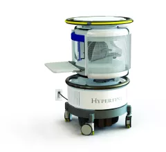
New miniature MRI sensor can detect electric currents, light in the brain

Massachusetts Institute of Technology (MIT) researchers have developed a minimally invasive MRI sensor that can detect electrical currents and light produced by luminescent proteins.

Philips unveils new radiation oncology portfolio for a more confident path to treatment at ASTRO 2018

New Big Bore RT and Ingenia Ambition/Elition MR-RT imaging systems and Pinnacle Evolution treatment planning software support improved patient care, accelerated time to treatment and enhanced patient satisfaction.

At ASTRO, Siemens Healthineers Debuts RT Pro Edition for MAGNETOM Sola for Aid in MRI Radiation Therapy Planning

At the 60th annual meeting of the American Society for Radiation Oncology (ASTRO), Oct. 21-24 at the Henry B. Gonzalez Convention Center in San Antonio, Siemens Healthineers unveils the MAGNETOM RT Pro edition for MAGNETOM Sola – a version of the company’s new 1.5 Tesla (1.5T) magnetic resonance imaging (MRI) scanner that is designed for assistance in radiation therapy (RT) treatment planning. In addition to BioMatrix technology, which addresses patients’ anatomical and physiological differences to overcome unwanted exam variability, the new scanner includes hardware and software that supports optimal treatment planning.

1.5T MRI scanner from Siemens Healthineers gains FDA clearance

The FDA has approved Siemens Healthineers’ MAGNETOM Sola, a 1.5 Tesla MRI scanner that utilizes the company's BioMatrix technology.

Thumbnail

MRI-based carotid artery measurements improve CVD risk assessment

MRI wall thickness measurements of a patient’s carotid arteries improves the quality of cardiovascular disease risk assessment, according to a new study published in Radiology.