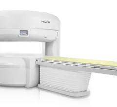
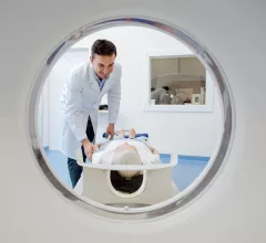
Magnetic Resonance Imaging

Magnetic resonance imaging (MRI) is the gold standard imaging modality for soft tissues. It produces detail cross-sectional images of soft tissue and bone anatomy, including muscles, tendons, ligaments, brain and organs, without the use of ionizing radiation. In addition to orthopedic imaging, MRI is also used for heart, brain and breast. MRI uses gadolinium contrast in many exams to highlight tissues and blood vessels, which enhances images and offers better diagnostic quality. It can also be used in conjunction with PET scans. How does MRI work? MR creates images by using powerful magnets to polarize hydrogen atoms in water (the body is made of of more than 80% water) so they face in one direction. A radiofrequency pulse is then used to ping these atoms, causing them to wobble, or resonate. The MRI coils detect this and computers can assemble images from the signals. Basic MRI scans will focus on the resonance of fat and water in two different sequences, which highlight and contrast different features in the anatomy.

Thumbnail

Diffusion MRI reveals memory capacity in healthy children

By assessing the degree of variegation in the white matter of children’s brains using diffusion-weighted MRI, neuroradiologists can predict how well the kids will perform in memory tests.

Thumbnail

When incidental renal lesions are discovered on lumbar spine MRI, what is the next step?

Incidental renal lesions are commonly detected during lumbar spine MRI examinations. When that occurs, is follow-up imaging always necessary? The authors of a new study published in the American Journal of Roentgenology explored that very question.

Thumbnail

Police filing new charges related to fatal MRI accident

Charges will be filed against ward boy Vitthal Chavan of Nair Hospital in Mumbai, India, for his involvement in the death of 32-year-old Rajesh Maru, who walked into an MRI room with a gas cylinder in January.

Thumbnail

MRI effective for pediatric appendicitis, even when read by adult-focused radiologists

Ultrasound (US) and CT are typically viewed as the most appropriate imaging modalities for diagnosing pediatric appendicitis, but what about MRI?

Thumbnail

10.5T MRI scanners tested on humans as imaging equipment keeps getting bigger

After four years of testing on animals and making key updates, the University of Minnesota’s Center for Magnetic Resonance Research in Minneapolis was finally ready to test its massive 10.5T MRI scanner on a human. And once the scan was complete, it turned out it was worth the wait.

Thumbnail

New technique brings MR fingerprinting to breast imaging

A new breast imaging technique uses MR fingerprinting for the volumetric quantification of T1 and T2 relaxation times for breast tissue, according to new research published in Radiology.

Thumbnail

FDA approves 7T MRI scanner at USC for clinical use

The 7T MRI scanner at the Keck School of Medicine of USC in Los Angeles has received FDA approval for clinical use.

Thumbnail

Installation of new MRI equipment leads to dozens of damaged iPhones, Apple Watches

It’s common knowledge that helium can make someone’s voice sound like a chipmunk, but the gas may be capable of another unexpected trick as well—doing serious damage to Apple products.