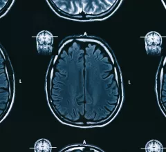
Magnetic Resonance Imaging

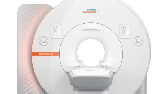
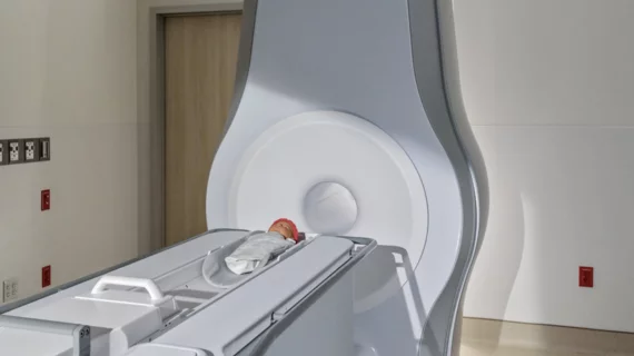
Magnetic resonance imaging (MRI) is the gold standard imaging modality for soft tissues. It produces detail cross-sectional images of soft tissue and bone anatomy, including muscles, tendons, ligaments, brain and organs, without the use of ionizing radiation. In addition to orthopedic imaging, MRI is also used for heart, brain and breast. MRI uses gadolinium contrast in many exams to highlight tissues and blood vessels, which enhances images and offers better diagnostic quality. It can also be used in conjunction with PET scans. How does MRI work? MR creates images by using powerful magnets to polarize hydrogen atoms in water (the body is made of of more than 80% water) so they face in one direction. A radiofrequency pulse is then used to ping these atoms, causing them to wobble, or resonate. The MRI coils detect this and computers can assemble images from the signals. Basic MRI scans will focus on the resonance of fat and water in two different sequences, which highlight and contrast different features in the anatomy.

Gas vesicles act as natural contrast agents, could result in 'erasable' MRIs

Scientists at the California Institute of Technology are taking steps toward erasable MRIs by using contrast agents natural to the human body—gas vesicles.

April 25, 2018
appendicitis acute abdomen

MR imaging a ‘suitable alternative’ to CT when evaluating patients for acute appendicitis

Physicians regularly turn to CT to evaluate patients who arrive at a hospital emergency department (ED) due to acute appendicitis. According to a new study published in Radiology, the diagnostic accuracy of MR imaging compares favorably to that of CT when diagnosing acute appendicitis, meaning patients can receive the same level of care without the ionizing radiation exposure.

April 25, 2018

Image reconstruction algorithm, MRI-derived heart strain values can aid prognosis in amyloidosis patients

Recent research found strain parameters taken from a cine MRI-based deformable registration algorithm (DRA) can determine the severity of amyloid buildup in the heart and may provide prognostic information on patients with light-chain (AL) amyloidosis.

April 24, 2018

New database measures safety of implantable devices in MR imaging

A streamlined database detailing protocol for scanning patients with a variety of implanted devices could be changing radiologists’ efficiency and confidence for the better, according to research out of Montefiore Medical Center in the Bronx, New York.

April 20, 2018
Waiting room

Machine learning model closely predicts patient waiting times for CT, MRI

Machine learning might be the next step in predicting patient wait times and appointment delays—factors crucial to healthcare’s quadruple aim and its emphasis on quality of care—in radiology practices, researchers have reported in the Journal of the American College of Radiology.

April 20, 2018

Canada notes 65% growth in MRI units across country

The same week British Columbia Health Minister Adrian Dix pledged $11 million to expand MRI usage across the Canadian province, the Canadian Agency for Drugs and Technologies in Health (CADTH) released its medical imaging inventory report for 2017, noting a 65 percent increase in MRI units in the past decade.

April 19, 2018

Should primary care physicians order MRIs for adult patients with knee pain?

When primary care physicians refer adult patients for MR imaging for traumatic knee symptoms, it provides no additional value and leads to higher healthcare costs, according to a new study by Dutch researchers published in Radiology.

April 17, 2018

8 surprising things people have done in MRI scanners—from opera to magic

MRI might be best used as a critical diagnostic tool for radiologists, but outside of the specialty, researchers have used the powerful machines to image patients’ brains while they do things from play video games to sing opera.

April 17, 2018