Magnetic Resonance Imaging

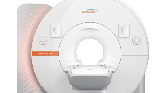
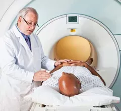
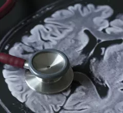
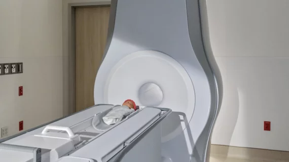
Magnetic resonance imaging (MRI) is the gold standard imaging modality for soft tissues. It produces detail cross-sectional images of soft tissue and bone anatomy, including muscles, tendons, ligaments, brain and organs, without the use of ionizing radiation. In addition to orthopedic imaging, MRI is also used for heart, brain and breast. MRI uses gadolinium contrast in many exams to highlight tissues and blood vessels, which enhances images and offers better diagnostic quality. It can also be used in conjunction with PET scans. How does MRI work? MR creates images by using powerful magnets to polarize hydrogen atoms in water (the body is made of of more than 80% water) so they face in one direction. A radiofrequency pulse is then used to ping these atoms, causing them to wobble, or resonate. The MRI coils detect this and computers can assemble images from the signals. Basic MRI scans will focus on the resonance of fat and water in two different sequences, which highlight and contrast different features in the anatomy.

Thumbnail

RSNA 2018: MRI scans reveal what football does to young athletes’ brains

Repeated blows to the head can cause changes to the brains of young football players, according to a new study presented at RSNA 2018 in Chicago.

Thumbnail

New Zealand-based promoter mandates pre-fight MRIs for all its boxers

Just weeks after charity fighter Kain Parsons was fatally injured in a Christchurch, New Zealand, boxing ring, sports promoter Duco Events has announced it’s implementing obligatory pre-fight MRIs for all of its boxers.

Thumbnail

NYU, Facebook release massive MRI dataset as part of ongoing AI project

The New York University (NYU) School of Medicine's department of radiology is releasing a knee MRI dataset of more than 1.5 million anonymous images as part of its ongoing collaboration with Facebook to make MRI scans 10 times faster with artificial intelligence (AI).

MRI scans using diffusion tensor imaging can predict Alzheimer's disease

MRI scans using diffusion tensor imaging (DTI) show potential for predicting if a patient will develop Alzheimer’s disease later in life, according to findings to be presented at RSNA 2018 in Chicago.

Thumbnail

AI startup Ezra secures $4M for MRI-based prostate cancer screening program

Ezra, a New York City-based artificial intelligence (AI) startup, has secured $4 million in funding for its new direct-to-consumer prostate cancer screening program.

Thumbnail

MRI captures more detail in colon cancers than multidetector CT

MRI may be a better choice than multidetector CT for identifying high-risk colon cancers that have already reached stage II or stage III, are still surgically resectable and are at risk of progressing to stage IV.

Thumbnail

MR spectroscopy adds little diagnostic value when imaging brain tumors

Adding magnetic resonance spectroscopy (MRS) to MRI does not significantly improve the classification of brain tumors in clinical practice, although MRS may be a valuable supplement to MRI in certain cases, according to researchers from Sweden's Uppsala University.

Thumbnail

Researcher awarded $40K to study how PET/MRI imaging could improve care for sarcoma patients

David Shultz, MD, PhD, of the Princess Margaret Cancer Centre in Toronto, Ontario, Canada, has been awarded the 2018 Hitachi Healthcare Americas /RSNA Research Seed Grant by the RSNA Research & Education (R&E) Foundation.