Magnetic Resonance Imaging

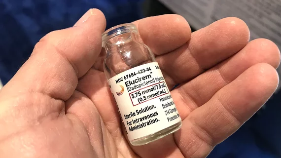
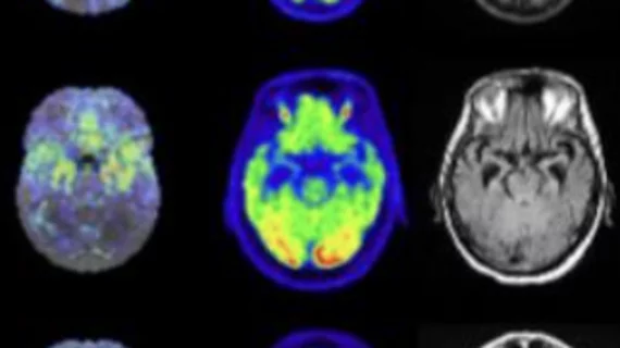
Magnetic resonance imaging (MRI) is the gold standard imaging modality for soft tissues. It produces detail cross-sectional images of soft tissue and bone anatomy, including muscles, tendons, ligaments, brain and organs, without the use of ionizing radiation. In addition to orthopedic imaging, MRI is also used for heart, brain and breast. MRI uses gadolinium contrast in many exams to highlight tissues and blood vessels, which enhances images and offers better diagnostic quality. It can also be used in conjunction with PET scans. How does MRI work? MR creates images by using powerful magnets to polarize hydrogen atoms in water (the body is made of of more than 80% water) so they face in one direction. A radiofrequency pulse is then used to ping these atoms, causing them to wobble, or resonate. The MRI coils detect this and computers can assemble images from the signals. Basic MRI scans will focus on the resonance of fat and water in two different sequences, which highlight and contrast different features in the anatomy.

Thumbnail

Breast MRI proves helpful with inconclusive digital mammography, DBT results

Radiologists can feel confident turning to breast MRI in instances when digital mammography (DM) and digital breast tomosynthesis (DBT) results are inconclusive, according to new research published in the American Journal of Roentgenology.

Thumbnail

Biannual MRI outperforms annual mammography for high-risk breast cancer patients

Biannual MRI is more effective than annual mammography in detecting early breast cancer in young women who have a high genetic risk of the disease, according to new research published in Clinical Cancer Research.

Thumbnail

New class of fractals could make for speedy whole-body MRI

An Australian researcher has said it could be possible to speed up full-body MRI by four times while controlling costs and maintaining quality—all thanks to the discovery of a new class of fractals. Shekhar Chandra, PhD, with the University of Queensland, believes “Chaotic Sensing,” an approach to sparse imaging using fractals, could help MRI machines to quickly identify necessary imaging information while discarding redundant data.
Thumbnail

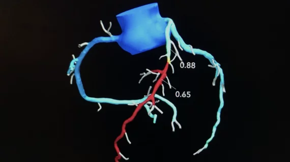
MRI helps build personalized 3D simulations of heart irregularities

Scientists hope personalized data from patients experiencing irregular heartbeats will help improve accuracy in heart ablation procedures. The 3D simulations of 21 patients allow physicians to locate arrhythmia by “poking” the simulated heart with small electrical signals in various locations.

Thumbnail

Varying prostate MRI insurance coverage poses problems for patients, providers

According to new research in the Journal of the American College of Radiology, prostate MRI has various problems—including varying insurance coverage, a failure to recognize major clinical scenarios and a restrictive, inflexible clinical practices.

Thumbnail

Physicians work to spread the word: MRI scans are generally safe for patients with pacemakers

While half a million of the U.K. population has pacemakers or defibrillators, a survey of England’s acute hospitals found that fewer than half would scan these cardiac patients using an MRI.

Thumbnail

Boston hospital installs first MRI system of its kind, says new 7T scanner now available

Brigham and Women’s Hospital (BWH) in Boston made two big imaging-related announcements this week. First, it has installed the first NICU-dedicated MRI system in the U.S. Second, its 7T scanner is now officially available for patients.

Thumbnail

Inner peace: MRI scans suggest mindful people may feel less pain

MRI scans show that more mindful individuals may feel less pain.