Magnetic Resonance Imaging

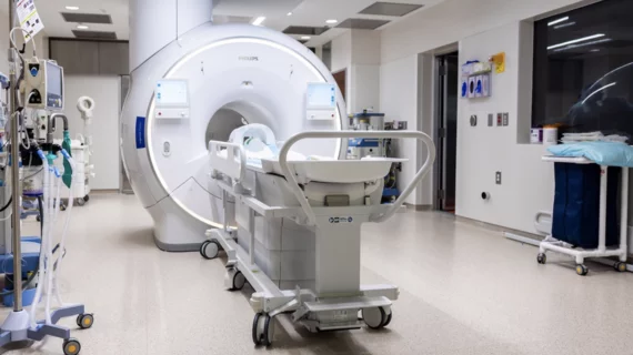
Magnetic resonance imaging (MRI) is the gold standard imaging modality for soft tissues. It produces detail cross-sectional images of soft tissue and bone anatomy, including muscles, tendons, ligaments, brain and organs, without the use of ionizing radiation. In addition to orthopedic imaging, MRI is also used for heart, brain and breast. MRI uses gadolinium contrast in many exams to highlight tissues and blood vessels, which enhances images and offers better diagnostic quality. It can also be used in conjunction with PET scans. How does MRI work? MR creates images by using powerful magnets to polarize hydrogen atoms in water (the body is made of of more than 80% water) so they face in one direction. A radiofrequency pulse is then used to ping these atoms, causing them to wobble, or resonate. The MRI coils detect this and computers can assemble images from the signals. Basic MRI scans will focus on the resonance of fat and water in two different sequences, which highlight and contrast different features in the anatomy.

Why do some women with dense breasts skip supplemental MRI after a negative mammogram?

Most women with extremely dense breasts who decline the invitation to receive supplemental MRI after a negative mammogram do so because of “MRI-related inconveniences” or anxiety, according to a new study published by Clinical Radiology.

May 15, 2018
anxiety-2987872_960_720.jpg

Research finds talking with radiographer calms patients before MRI

Nerves before an MRI are normal—up to 37 percent of patients report either moderate or high levels of anxiety leading up to an exam—and this apprehension can have physical consequences that render an entire scanning experience useless, first author J.R. Tugwell and colleagues wrote in Radiography this month.

May 14, 2018
Cheryl Petersilge, MD, MBA, with the department of regional radiology at the Cleveland Clinic, examined enterprise imaging—and how radiologists must integrate and collaborate with other departments. Her clinical perspective clinical perspective was published online in the October issue of the American Journal of Roentgenology.

Extracting radiomic features from MR images helps with breast lesion classification

Extracting radiomic features from MR images can help radiologists distinguish between benign breast lesions and luminal A breast cancers, according to a new study published by Academic Radiology.

May 14, 2018

Expensive imaging: New data reveal MRI prices highest in the US

The average price of an MRI in the United States is $1,119, higher than in any comparable countries, according to a new report from the Kaiser Family Foundation. The report examined the availability, utilization and prices of a variety of health services in countries all over the world.

May 11, 2018

Multicenter trial finds MRI-targeted biopsies superior to standard biopsies for diagnosing prostate cancer

When treating men with a clinical suspicion of prostate cancer, providers often turn to an ultrasound-guided biopsy of the prostate, though that method has been associated with missing clinically significant cancers and detecting clinically insignificant cancers.

May 10, 2018

Patients in yoga pants suffering burns during MRIs

If patients aren’t dressed appropriately when they undergo MRIs, they can suffer significant burns during the procedure. Due to small metal threads in the material, yoga pants and other similar clothing are a becoming a prime culprit.

May 9, 2018
screen_shot_2018-05-09_at_10.30.13_am.png

Wearable MRI glove captures clear images of bones, tendons moving together

A novel, flexible MRI component hit the national stage this week in the form of a glove, New York University researchers report. It is delivering science’s first-ever clear images of bones, tendons and ligaments moving together.

May 9, 2018

ABVS means improved comfort, satisfaction for patients who would otherwise undergo MRI

Automated Breast Volume Scanner (ABVS) evaluation is close to matching traditional breast MRI in assessing tumor diameter and volume—and it’s leaving patients more satisfied and comfortable than its conventional counterpart, a study published in the European Journal of Radiology suggests.

May 8, 2018