Magnetic Resonance Imaging

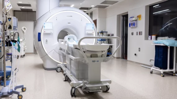
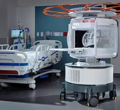
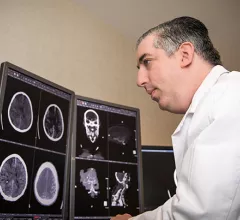
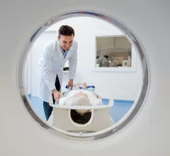
Magnetic resonance imaging (MRI) is the gold standard imaging modality for soft tissues. It produces detail cross-sectional images of soft tissue and bone anatomy, including muscles, tendons, ligaments, brain and organs, without the use of ionizing radiation. In addition to orthopedic imaging, MRI is also used for heart, brain and breast. MRI uses gadolinium contrast in many exams to highlight tissues and blood vessels, which enhances images and offers better diagnostic quality. It can also be used in conjunction with PET scans. How does MRI work? MR creates images by using powerful magnets to polarize hydrogen atoms in water (the body is made of of more than 80% water) so they face in one direction. A radiofrequency pulse is then used to ping these atoms, causing them to wobble, or resonate. The MRI coils detect this and computers can assemble images from the signals. Basic MRI scans will focus on the resonance of fat and water in two different sequences, which highlight and contrast different features in the anatomy.

Untraditional teaching tool underscores importance of MRI safety for radiology trainees

Applying an alternative deep learning, assessment-based approach to radiology education could help trainees better retain information for use in future work, according to a study published in Radiography this month.

June 15, 2018

Can biparametric MRI accurately detect prostate cancer?

Research from the last 17 years shows that biparametric MRI (bpMRI) gives radiologists an accurate tool for detecting prostate cancer (PCa), according to a new meta-analysis published by the American Journal of Roentgenology.

June 14, 2018

MRI finds brains of children hooked on video games similar to those of drug, alcohol addicts

An MRI study out of California has found excessive social media and addictive video games can have an effect on children’s brains similar to that of drug abuse or alcoholism.

June 14, 2018

Arterial spin labeling MRI explains cognitive dysfunction in young kidney disease patients

Arterial spin labeling MRI may offer a noninvasive alternative for quantifying cerebral blood flow without the use of contrast agents—a necessity for patients with illnesses like kidney disease, researchers wrote in a Radiology study this June.

June 13, 2018

MRI adds value to endometrial cancer prognosis

Including MRI in the routine evaluation of endometrial cancer could help assess the aggressiveness of tumors while stratifying which patients may benefit from surgeries like lymphadenectomy, researchers reported in Clinical Radiology this June.

June 11, 2018
draw-2103019_960_720.jpg

Auburn launches interdisciplinary MRI trial to study brain plasticity

A $90,000 grant from the National Endowment for the Arts is offering Auburn University imagers an opportunity to study the effect art classes can have on brain plasticity, the university announced this week. Results could have important implications for how cognitive impairment cases are handled.

June 11, 2018

Researchers develop rice-sized ‘seed’ that destroys deadly brain tumors with the help of MRI

A magnetic metal “seed,” no larger than a grain of rice and developed by scientists at University College London, has the ability to rapidly kill cancer cells and destroy deadly tumors within 10 minutes, the Telegraph reported this week.

June 8, 2018

India debuts its first-ever portable MRI scanner

Voxelgrids, an offshoot of India’s Tata Trusts Foundation for Innovation and Social Entrepreneurship, announced June 7 its latest development: a lightweight, portable MRI scanner that could cut imaging costs in half across the country. 

June 7, 2018