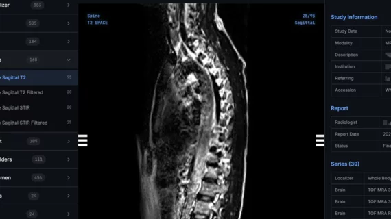
Magnetic Resonance Imaging

Magnetic resonance imaging (MRI) is the gold standard imaging modality for soft tissues. It produces detail cross-sectional images of soft tissue and bone anatomy, including muscles, tendons, ligaments, brain and organs, without the use of ionizing radiation. In addition to orthopedic imaging, MRI is also used for heart, brain and breast. MRI uses gadolinium contrast in many exams to highlight tissues and blood vessels, which enhances images and offers better diagnostic quality. It can also be used in conjunction with PET scans. How does MRI work? MR creates images by using powerful magnets to polarize hydrogen atoms in water (the body is made of of more than 80% water) so they face in one direction. A radiofrequency pulse is then used to ping these atoms, causing them to wobble, or resonate. The MRI coils detect this and computers can assemble images from the signals. Basic MRI scans will focus on the resonance of fat and water in two different sequences, which highlight and contrast different features in the anatomy.

Thumbnail

What we can learn about stroke from a simple MRI technique

Using MRI scans to measure iron content can help specialists learn more about stroke-related damage to the brain, according to a new study published by Radiology. Should such measurements be required after patients suffer a stroke?

Thumbnail

Second-opinion reviews of breast MRI studies provide value

Second-opinion breast MRI reviews by subspecialized radiologists can improve patient management and increase cancer detection, according to findings published in the Journal of the American College of Radiology.

Thumbnail

No gadolinium, no problem: Specialists can monitor MS patients with unenhanced MRI

Gadolinium-based contrast agents (GBCAs) are typically administered during follow-up imaging of multiple sclerosis (MS) patients. According to new research published in Radiology, however, unenhanced MRI scans are sufficient.

Thumbnail

Should pediatric patients be excluded from MRI research due to stress, anxiety? 

Children and teenagers undergoing brain MRI scans experience little to no more claustrophobia or other form of anxiety than adults, so there’s no reason to exclude pediatric patients from MRI research on the basis of concern over stress. 

Thumbnail

How custom worklists can lead to faster MRI interpretation times

Analytics-driven worklists can help entire groups of radiologists achieve faster MRI interpretation times, according to new research published in the American Journal of Roentgenology.

Thumbnail

Experience not an advantage when radiologists read prostate MRI

In a recently completed study at Washington University Medical Center in St. Louis, experienced radiologists were no more accurate than younger colleagues at diagnosing prostate cancer on multiparametric MRI.

Siemens Healthineers Announces First U.S. Installation of MAGNETOM Sola 1.5T MRI Scanner

South Texas Radiology Imaging Centers, San Antonio, recently became the first healthcare institution in the United States to install the MAGNETOM Sola, a 1.5 Tesla (1.5T), 70-cm magnetic resonance imaging (MRI) scanner from Siemens Healthineers with a new magnet design in addition to BioMatrix patient personalization technology.

Thumbnail

Simple solution emerges on arm MRI for obese, claustrophobic patients

Imagers challenged to complete arm MR scans for extremely claustrophobic or morbidly obese patients can do the job with a standard scanner, a wrist and elbow coil—and enough space behind the scanner for the patient to sit or stand alongside.