Magnetic Resonance Imaging

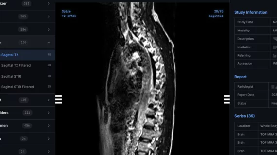
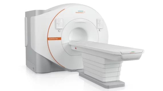
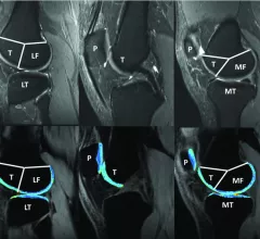
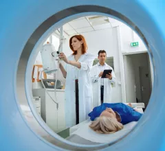
Magnetic resonance imaging (MRI) is the gold standard imaging modality for soft tissues. It produces detail cross-sectional images of soft tissue and bone anatomy, including muscles, tendons, ligaments, brain and organs, without the use of ionizing radiation. In addition to orthopedic imaging, MRI is also used for heart, brain and breast. MRI uses gadolinium contrast in many exams to highlight tissues and blood vessels, which enhances images and offers better diagnostic quality. It can also be used in conjunction with PET scans. How does MRI work? MR creates images by using powerful magnets to polarize hydrogen atoms in water (the body is made of of more than 80% water) so they face in one direction. A radiofrequency pulse is then used to ping these atoms, causing them to wobble, or resonate. The MRI coils detect this and computers can assemble images from the signals. Basic MRI scans will focus on the resonance of fat and water in two different sequences, which highlight and contrast different features in the anatomy.

Thumbnail

Fast MRI scans ID brain injuries in young patients without radiation, sedation

“Fast MRI” scans, which use quicker imaging techniques and don’t require sedation or ionizing radiation, can identify traumatic brain injuries (TBIs) in young patients, according to a new study published in Pediatrics.

Thumbnail

New MRI technique predicts dementia in stroke patients

A new MRI technique using diffusion tensor imaging (DTI) can help predict when patients might suffer from stroke-related dementia, according to new research published in Stroke: A Journal of Cerebral Circulation.

Thumbnail

New MRI technique tracks molecular changes in the brain as it ages

A team of researchers has found that quantitative MRI (qMRI) can be used to monitor molecular changes in the brain, sharing its findings in Nature Communications.

Thumbnail

New MRI technique measures heart strain without gadolinium

Researchers have developed a new MRI technique that allows providers to measure strain in heart muscles without the use of gadolinium, according to new findings published in Scientific Reports.

Thumbnail

ACR publishes new guidance on prostate MRI utilization

The American College of Radiology (ACR) has developed a new guidance document on the use of prostate MRI for working up suspected or known malignancies.

Thumbnail

AI techniques detect implantable devices, improve MRI safety

Two different AI-based methods can identify patients with implantable devices that could pose a safety risk during MRI scans, according to research published in the Journal of the American College of Radiology.

FDA approves use of Boston Scientific DBS system during full-body MRI scans

Boston Scientific Corporation announced that it has received FDA approval for its Vercise Gevia deep brain stimulation (DBS) system to be used during full-body MRI scans.

Thumbnail

Gadolinium-free contrast agent could improve MRI safety

A new manganese-based contrast agent could help limit the use of gadolinium-based contrast agents (GBCAs) and make MRI scans safer, according to findings published in Investigative Radiology.