Magnetic Resonance Imaging

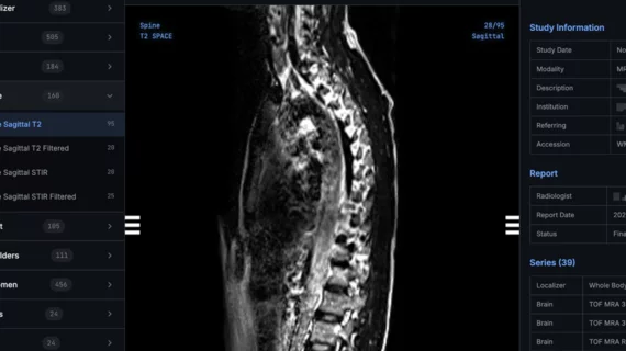
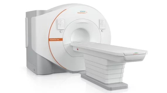
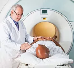
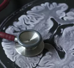
Magnetic resonance imaging (MRI) is the gold standard imaging modality for soft tissues. It produces detail cross-sectional images of soft tissue and bone anatomy, including muscles, tendons, ligaments, brain and organs, without the use of ionizing radiation. In addition to orthopedic imaging, MRI is also used for heart, brain and breast. MRI uses gadolinium contrast in many exams to highlight tissues and blood vessels, which enhances images and offers better diagnostic quality. It can also be used in conjunction with PET scans. How does MRI work? MR creates images by using powerful magnets to polarize hydrogen atoms in water (the body is made of of more than 80% water) so they face in one direction. A radiofrequency pulse is then used to ping these atoms, causing them to wobble, or resonate. The MRI coils detect this and computers can assemble images from the signals. Basic MRI scans will focus on the resonance of fat and water in two different sequences, which highlight and contrast different features in the anatomy.

Thumbnail

Improved radiology reports, clinician education improve knee MRI utilization

Researchers have found that standardized radiology reports and clinician education can reduce unnecessary knee MRI utilization for patients with severe osteoarthritis (OA).

Thumbnail

AI model predicts a patient’s breast cancer risk from single MR image

Researchers have developed a deep learning (DL) model that assesses a woman’s five-year cancer risk with a single breast MR image, sharing their findings in the American Journal of Roentgenology.

Thumbnail

Both major parties in Australia commit to funding MRI scans for breast cancer patients

The two major parties set to take part in Australia’s upcoming federal election have both committed to increase funding for MRI scans for women with breast cancer.

Thumbnail

7T MRI scans improve care for patients with focal epilepsy

7T MRI scans of patients with focal epilepsy can provide valuable information missed by 3T scans, according to new research published in PLOS ONE.

Thumbnail

Updated MRI protocol reduces need to sedate pediatric patients

An abbreviated brain MRI protocol can help reduce the need to sedate young children suffering from headaches, according to a new study published by the American Journal of Roentgenology.

Thumbnail

What we can learn about stroke from a simple MRI technique

Using MRI scans to measure iron content can help specialists learn more about stroke-related damage to the brain, according to a new study published by Radiology. Should such measurements be required after patients suffer a stroke?

Thumbnail

Second-opinion reviews of breast MRI studies provide value

Second-opinion breast MRI reviews by subspecialized radiologists can improve patient management and increase cancer detection, according to findings published in the Journal of the American College of Radiology.

Thumbnail

No gadolinium, no problem: Specialists can monitor MS patients with unenhanced MRI

Gadolinium-based contrast agents (GBCAs) are typically administered during follow-up imaging of multiple sclerosis (MS) patients. According to new research published in Radiology, however, unenhanced MRI scans are sufficient.