Magnetic Resonance Imaging

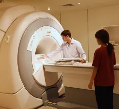
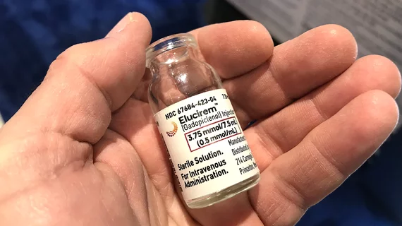
Magnetic resonance imaging (MRI) is the gold standard imaging modality for soft tissues. It produces detail cross-sectional images of soft tissue and bone anatomy, including muscles, tendons, ligaments, brain and organs, without the use of ionizing radiation. In addition to orthopedic imaging, MRI is also used for heart, brain and breast. MRI uses gadolinium contrast in many exams to highlight tissues and blood vessels, which enhances images and offers better diagnostic quality. It can also be used in conjunction with PET scans. How does MRI work? MR creates images by using powerful magnets to polarize hydrogen atoms in water (the body is made of of more than 80% water) so they face in one direction. A radiofrequency pulse is then used to ping these atoms, causing them to wobble, or resonate. The MRI coils detect this and computers can assemble images from the signals. Basic MRI scans will focus on the resonance of fat and water in two different sequences, which highlight and contrast different features in the anatomy.

Thumbnail

Should pediatric patients be excluded from MRI research due to stress, anxiety? 

Children and teenagers undergoing brain MRI scans experience little to no more claustrophobia or other form of anxiety than adults, so there’s no reason to exclude pediatric patients from MRI research on the basis of concern over stress. 

Thumbnail

How custom worklists can lead to faster MRI interpretation times

Analytics-driven worklists can help entire groups of radiologists achieve faster MRI interpretation times, according to new research published in the American Journal of Roentgenology.

Thumbnail

Experience not an advantage when radiologists read prostate MRI

In a recently completed study at Washington University Medical Center in St. Louis, experienced radiologists were no more accurate than younger colleagues at diagnosing prostate cancer on multiparametric MRI.

Siemens Healthineers Announces First U.S. Installation of MAGNETOM Sola 1.5T MRI Scanner

South Texas Radiology Imaging Centers, San Antonio, recently became the first healthcare institution in the United States to install the MAGNETOM Sola, a 1.5 Tesla (1.5T), 70-cm magnetic resonance imaging (MRI) scanner from Siemens Healthineers with a new magnet design in addition to BioMatrix patient personalization technology.

Thumbnail

Simple solution emerges on arm MRI for obese, claustrophobic patients

Imagers challenged to complete arm MR scans for extremely claustrophobic or morbidly obese patients can do the job with a standard scanner, a wrist and elbow coil—and enough space behind the scanner for the patient to sit or stand alongside.

Thumbnail

AI IDs lung tumors from MRI scans better than other methods

A new AI algorithm developed by researchers at the University of Alberta in Edmonton, Alberta, Canada, can identify lung tumors from MRI scans faster than other advanced methods.

Thumbnail

Cardiac MRI-derived T2 mapping may help heart failure patients

Researchers used T2 mapping taken from weekly cardiac MRIs to help identify cardiotoxicity at an early stage, according to results of a pig study published Feb. 18 in the Journal of the American College of Cardiology. The findings could help cancer patients at risk of chemo-induced heart failure.

Thumbnail

Breast imaging referrers ordering more MRI scans following legislation

At an academic health system in Boston, referring physicians have been ordering more breast MRI scans since Massachusetts mandated that mammographers inform patients in writing of their breast density.