Magnetic Resonance Imaging

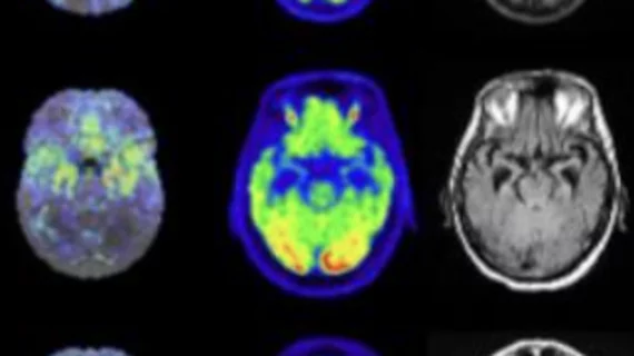
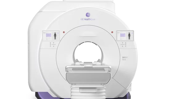
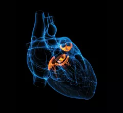
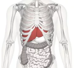
Magnetic resonance imaging (MRI) is the gold standard imaging modality for soft tissues. It produces detail cross-sectional images of soft tissue and bone anatomy, including muscles, tendons, ligaments, brain and organs, without the use of ionizing radiation. In addition to orthopedic imaging, MRI is also used for heart, brain and breast. MRI uses gadolinium contrast in many exams to highlight tissues and blood vessels, which enhances images and offers better diagnostic quality. It can also be used in conjunction with PET scans. How does MRI work? MR creates images by using powerful magnets to polarize hydrogen atoms in water (the body is made of of more than 80% water) so they face in one direction. A radiofrequency pulse is then used to ping these atoms, causing them to wobble, or resonate. The MRI coils detect this and computers can assemble images from the signals. Basic MRI scans will focus on the resonance of fat and water in two different sequences, which highlight and contrast different features in the anatomy.

Thumbnail

Patient at Massachusetts hospital breaks facial bone after ‘freak accident’ in MRI suite

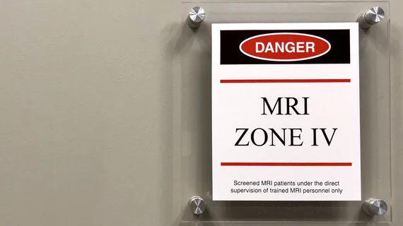
An MRI patient in Lowell, Massachusetts, sustained a facial fracture last month after a technologist at Lowell General Hospital Saints Campus introduced a metal hamper to the room, causing the hamper to fly toward the MRI’s magnet and strike the man in his face, the Lowell Sun reported.

Thumbnail

The evolution of the EMR: Radiologists reveal how they’d react to a new system

Radiology leaders responded this week to a difficult hypothetical—something the Journal of the American College of Radiology itself called a “devilish dilemma”: What would they do if their hospitals insisted on permanently switching to a new electronic medical record (EMR) without first consulting them?

Thumbnail

AHRA 2018: Tobias Gilk on why imaging providers should take MRI safety more seriously

Tobias Gilk, MArch, MRSO, MRSE, has made it his personal mission to improve MRI safety in the United States. Gilk spoke with Radiology Business at the AHRA 2018 Annual Meeting in Orlando to discuss that mission.

Thumbnail

Could this MRI technique help predict disabilities in multiple sclerosis patients?

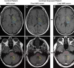
Researchers have shown that a new MRI technique can measure brain iron levels in multiple sclerosis (MS) patients, according to a study published in Radiology. This breakthrough can help identify MS patients at an especially high risk of developing physical disabilities.

Massachusetts county's MRIs offer more bang for the out-of-pocket buck

Worcester County, Massachusetts, residents can enjoy a better deal on MRIs than anywhere else in the state, the Telegram & Gazette reported of a Pioneer Institute study released this week, averaging out-of-pocket costs that rarely exceed $60.

Thumbnail

MRI research inspires mayor to propose new necktie policy

R. Rex Parris, the mayor of Lancaster, California, has proposed the city ban local employees from being required to wear a necktie. The decision, he said, was made after reading about recent imaging research that found neckties can lead to a reduction in cerebral blood flow.

Thumbnail

Machine learning analyzes MRIs to identify schizophrenia with 78% accuracy

Researchers have shown that machine learning can identify if a patient has schizophrenia by analyzing an MRI of their brain, according to a new study published in Molecular Psychiatry.

Thumbnail

Multiparametric MRI an effective tool for monitoring prostate cancer patients after focal therapy

Multiparametric MRI can provide value by highlighting changes in the prostate following MRI-guided focal laser ablation (FLA), according to a new study published by the American Journal of Roentgenology.