Molecular Imaging

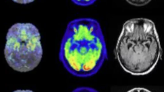
Molecular imaging (also called nuclear medicine or nuclear imaging) can image the function of cells inside the body at the molecular level. This includes the imaging modalities of positron emission computed tomography (PET) and single photon emission computed tomography (SPECT) imaging. How does PET and SPECT imaging work? Small amounts of radioactive material (radiopharmaceuticals) injected into a patient. These can use sugars or chemical traits to bond to specific cells. The radioactive material is taken up by cells that consume the sugars. The radiation emitted from inside the body is detected by photon detectors outside the body. Computers take the data to assemble images of the radiation emissions. Nuclear images may appear fuzzy or ghostly rather than the sharper resolution from MRI and CT.  But, it provides metabolic information at a cellular level, showing if there are defects in the function of the heart, areas of very high metabolic activity associated with cancer cells, or areas of inflammation, data not available from other modalities. These noninvasive imaging exams are used to diagnose cancer, heart disease, Alzheimer’s and Parkinson’s disease, bone disorders and other disorders. 

Thumbnail

MRI enhances cellular resolution to determine effectiveness of cancer therapy

Researchers from the Keck School of Medicine at the University of Southern California (USC) successfully used high-resolution MRI and a cell-labeling technique to visualize nearly 100 cells, which may allow clinicians to assess the effectiveness of immune cell- and stem cell-based therapies to treat cancer.

Thumbnail

Can a new clinical state for prostate cancer help personalize treatment for the disease?

For patients with metastatic prostate cancer, understanding its genesis and evolution can mean all the difference for treatment. But, what if, as a recent Journal of Nuclear Medicine perspective proposes, a new “clinical state” could improve understanding and treatment of the disease?

Thumbnail

SNMMI, imaging leaders back $20M in federal funds for Mo-99 production

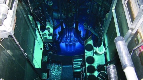
The Society for Nuclear Medicine and Molecular Imaging (SNMMI), along with a host of other leading medical imaging groups and vendors, addressed a July 18 letter to the Senate and House subcommittees on Energy and Water Development voicing its support for $20 million in additional funding to help establish domestic production of molybdenum-99 (Mo-99).

Thumbnail

PET imaging helps map Parkinson’s damage to heart

A team from the University of Wisconsin-Madison successfully used PET imaging to visualize heart damage commonly seen in those with Parkinson’s disease. The technique will help develop therapies.

Thumbnail

Rural patients suffer as Tc-99m production stops at Australia's only nuclear medicine generator

The shutdown of Australia’s sole nuclear medicine generator due to mechanical problems is beginning to impact patient treatment, particularly for individuals in rural parts of the country, the Guardian reported.

Thumbnail

Molecular imaging becomes 3D interactive experience with VR

A team of researchers has created a virtual reality (VR) environment that allows users to manipulate 3D molecular proteins 10 times faster than with a 2D screen, according to a July 3 article by the New York Times.

Thumbnail

Fluciclovine PET/CT IDs recurrent prostate cancer, alters patient management

Adding fluorine-18 (18F)-fluciclovine PET/CT to the diagnostic treatment plan of patients with biochemical recurrence of prostate cancer can identify previously undetected lesions and alter treatment management for patients with the disease.

Thumbnail

MR-assisted PET data optimization may improve neuroanalysis of dementia patients

Researchers—led by Kevin Chen, PhD, a postdoctoral fellow at Harvard University's Microrobotics Lab—have found that spatiotemporally correlated data acquired using a single magnetic resonance (MR) sequence may be successfully used in a PET/MRI scanner for PET attenuation, motion and partial effects corrections.