Molecular Imaging

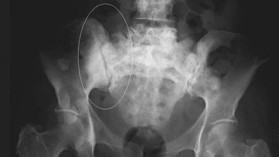
Molecular imaging (also called nuclear medicine or nuclear imaging) can image the function of cells inside the body at the molecular level. This includes the imaging modalities of positron emission computed tomography (PET) and single photon emission computed tomography (SPECT) imaging. How does PET and SPECT imaging work? Small amounts of radioactive material (radiopharmaceuticals) injected into a patient. These can use sugars or chemical traits to bond to specific cells. The radioactive material is taken up by cells that consume the sugars. The radiation emitted from inside the body is detected by photon detectors outside the body. Computers take the data to assemble images of the radiation emissions. Nuclear images may appear fuzzy or ghostly rather than the sharper resolution from MRI and CT.  But, it provides metabolic information at a cellular level, showing if there are defects in the function of the heart, areas of very high metabolic activity associated with cancer cells, or areas of inflammation, data not available from other modalities. These noninvasive imaging exams are used to diagnose cancer, heart disease, Alzheimer’s and Parkinson’s disease, bone disorders and other disorders. 

Thumbnail

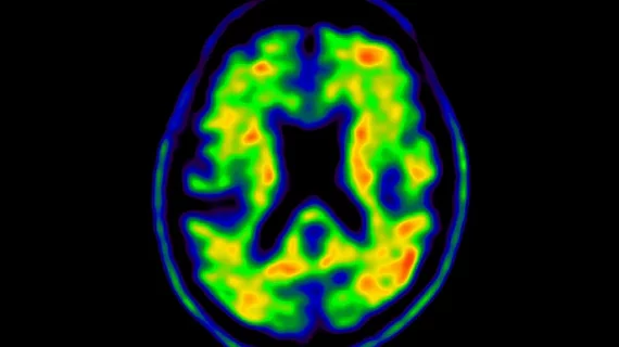
Amyloid load is a more sensitive biomarker for Alzheimer’s imaging

Amyloid load (AβL) imaged in vivo using [18F]Florbetapir PET is a more sensitive biomarker for measuring the global amyloid (Aβ) burden compared to composite standardized uptake value ratio (SUVr), reported authors of a Sept. 6 Journal of Nuclear Medicine study.

Thumbnail

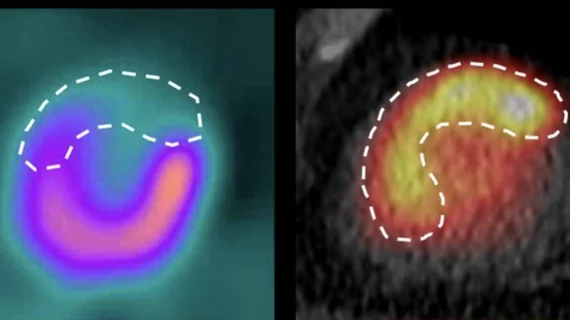
SNMMI supports proposed expansion of cardiac PET coverage

The Society of Nuclear Medicine and Molecular Imaging (SNMMI) and the American Society of Nuclear Cardiology (ASNC) recently submitted comment to BlueCross BlueShield (BCBS) of Tennessee supporting its proposed expansion of cardiac PET coverage.

Thumbnail

Experts find flaw in prostate cancer PET imaging technique

Authors of a recent Journal of Nuclear Medicine study have discovered potential for misdiagnosis when relying solely on prostate-specific membrane antigen (PSMA) PET imaging in prostate cancer staging.

Thumbnail

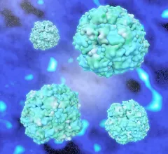
Electron microscopic imaging of virus may reveal new potential for gene therapy

Researchers from the Salk Institute in San Diego and the University of Florida have used cryo-electron microscopy (cryo-EM) imaging to analyze a 3D model of the AAV2 virus. The advanced molecular imaging technique may demonstrate the potential for the virus to act as a delivery vehicle for gene therapies, according to research published Sept. 7 in Nature Communications.

Thumbnail

Novel nuclear imaging method targets cancer-associated fibroblasts, outperforms 18F-FDG

A German-led research team has developed a new nuclear medicine imaging technique that outperforms standard tumor imaging by targeting cancer-associated fibroblasts (CAFs), according to research published in the September issue of the Journal of Nuclear Medicine.

Thumbnail

Amyloid PET improves diagnosis, treatment in Alzheimer’s patients

For patients with Alzheimer’s disease, amyloid PET imaging can improve diagnosis, diagnostic confidence and treatment, according to a JAMA Neurology study. The method is beneficial whether results are positive or negative.

Aetna expands coverage for Ga-68 dotatate PET

Aetna announced, Thursday, Aug. 16, it will expand its coverage of the PET tracer gallium-68 (Ga-68) dotatate, according to a Society of Nuclear Medicine and Molecular Imaging (SNMMI) release. The radiopharmaceutical is now considered “medically necessary.”

Thumbnail

15-minute PET acquisition time for rectal cancer improves care—without extending scan time

Extending PET acquisition time during PET/MRI for rectal cancer staging increases identified lymph nodes and has no impact on scan time, reported authors of a recent American Journal of Roentgenology study.