Molecular Imaging

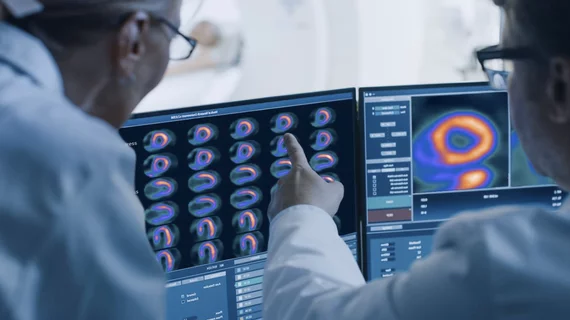
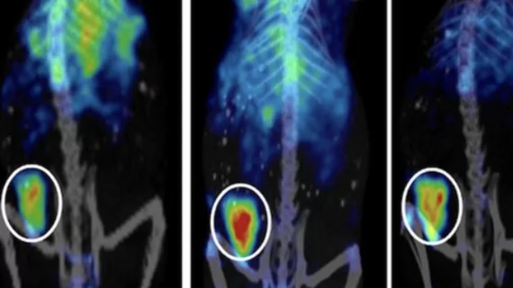
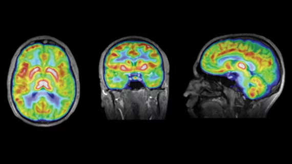
Molecular imaging (also called nuclear medicine or nuclear imaging) can image the function of cells inside the body at the molecular level. This includes the imaging modalities of positron emission computed tomography (PET) and single photon emission computed tomography (SPECT) imaging. How does PET and SPECT imaging work? Small amounts of radioactive material (radiopharmaceuticals) injected into a patient. These can use sugars or chemical traits to bond to specific cells. The radioactive material is taken up by cells that consume the sugars. The radiation emitted from inside the body is detected by photon detectors outside the body. Computers take the data to assemble images of the radiation emissions. Nuclear images may appear fuzzy or ghostly rather than the sharper resolution from MRI and CT.  But, it provides metabolic information at a cellular level, showing if there are defects in the function of the heart, areas of very high metabolic activity associated with cancer cells, or areas of inflammation, data not available from other modalities. These noninvasive imaging exams are used to diagnose cancer, heart disease, Alzheimer’s and Parkinson’s disease, bone disorders and other disorders. 

Thumbnail

Researches identify features least-sensitive to PET system variations

Researchers from the Medical University of Vienna in Austria provided new guidance for selecting optimizing features from 18F-FDG-PET/CT studies—demonstrating feature variations can be minimized for selected image parameters and imaging systems, in a new study published Nov. 2 in the Journal of Nuclear Medicine.

Thumbnail

SNMMI issues alert over expected Mo-99 shortage

The Society of Nuclear Medicine and Molecular Imaging (SNMMI) issued an alert Wednesday, Oct. 31, warning members of an expected shortage of Mo-99 during the first-half of November, according to a news release.

Thumbnail

Clinical trial involving novel PET agent may help Parkinson’s patients

An upcoming study will test a new imaging agent which allows visualization of human synapses, according to a news release. Results will inform a future trial designed to aid Parkinson’s patients.

Thumbnail

Communication is key: Nuclear medicine departments fail diabetes patients preparing for 18-FDG PET/CT

Nuclear medicine departments are not communicating proper pre-scan instructions to diabetes mellitus management patients for optimizing fluoride-18 fluorodeoxyglucose (FDG) PET/CT imaging, according to new research published in the Journal of the American College of Radiology.

PET/CT diagnoses blood vessel disease better than biopsy method

Pairing PET with CT achieved better diagnostic accuracy than temporal artery biopsy (TAB) in patients with newly suspected giant cell arteritis (GCA), according to research presented recently at the 2018 American College of Rheumatology (ACR)/ Association of Reproductive Health Professional (AHRP) meeting in Chicago.

Thumbnail

Novel molecular CT technique could revolutionize drug discovery

The technique is able to map the structure of small organic molecules, such as pharmaceuticals and hormones, with electron diffraction imaging—commonly used to chart larger proteins, according to a report published Oct. 19 by Science Magazine.

Thumbnail

PET quantification method can help improve amyloid pathology assessment

Utilizing non-displaceable binding potential (BPND) quantification with 18F-Flutemetamol PET imaging can better assess amyloid pathology in elderly patients with low amyloid buildup compared to traditional standard uptake value ratio (SUVr), reported authors of an Oct. 12 study in the Journal of Nuclear Medicine.

Thumbnail

PET can image T-cells for chronic liver disease

PET imaging could serve as an accurate, noninvasive substitute to liver biopsies in patients with chronic liver diseases, as detailed in research published in the October issue of The Journal of Nuclear Medicine.