Molecular Imaging

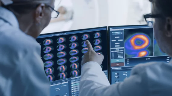
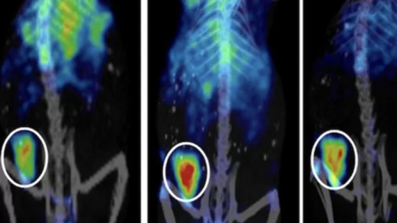
Molecular imaging (also called nuclear medicine or nuclear imaging) can image the function of cells inside the body at the molecular level. This includes the imaging modalities of positron emission computed tomography (PET) and single photon emission computed tomography (SPECT) imaging. How does PET and SPECT imaging work? Small amounts of radioactive material (radiopharmaceuticals) injected into a patient. These can use sugars or chemical traits to bond to specific cells. The radioactive material is taken up by cells that consume the sugars. The radiation emitted from inside the body is detected by photon detectors outside the body. Computers take the data to assemble images of the radiation emissions. Nuclear images may appear fuzzy or ghostly rather than the sharper resolution from MRI and CT.  But, it provides metabolic information at a cellular level, showing if there are defects in the function of the heart, areas of very high metabolic activity associated with cancer cells, or areas of inflammation, data not available from other modalities. These noninvasive imaging exams are used to diagnose cancer, heart disease, Alzheimer’s and Parkinson’s disease, bone disorders and other disorders. 

Thumbnail

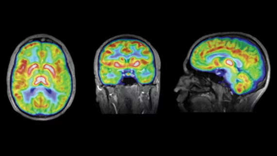
Automated amyloid PET quantification approach can help ID Alzheimer’s

Using an automated technique to quantify amyloid plaque on PET scans in a patient’s native space can improve the detection of brain amyloid accumulation compared to traditional methods, according to a recent study published in the Journal of Nuclear Medicine.

Thumbnail

Australia’s $168M nuclear medicine plant begins full production of Mo-99

Until now, the $168 million facility was producing limited amounts of Mo-99, a radioisotope used in approximately 85% of all Australian nuclear medicine procedures such as SPECT scans.

Thumbnail

Radiotracer identifies 28 different forms of cancer

A team of German researchers has found a new class of radiopharmaceuticals capable of identifying 28 types of malignant tumors, imaging them with high uptake and image contrast.


Thumbnail

USP publishes new safety standards for radiopharmaceuticals

The U.S. Pharmacopeia (USP) recently published a new chapter outlining minimum standards for preparing, compounding, dispensing and packaging sterile and non-sterile radiopharmaceuticals that are part of state-licensed activities.

Breakthrough method may improve cost, efficiency of radioisotope production

The approach uses a linear electron accelerator and naturally-occurring molybdenum-100, eliminating the need for a nuclear reactor or enriched uranium.

Thumbnail

SHINE gains exclusive license for Lu-177 radioisotope production

Janesville, Wisconsin-based SHINE Medical Technologies has announced an agreement with the Institute of Organic Chemistry and Biochemistry of the CAS (IOCB Prague), gaining exclusive rights over an innovation to produce lutetium-177 (Lu-177).

Thumbnail

Novel imaging agent utilizes protein found in scorpion venom

Researchers have combined a new imaging technique and imaging agent which utilizes a synthesized amino acid found in scorpion venom to help illuminate brain tumors during surgery.

Thumbnail

US Nuclear Regulatory Commission seeks feedback on radiopharmaceutical training requirements

The U.S. Nuclear Regulatory Commission (NRC) opened a 30-day public comment period seeking feedback on its draft document outlining training and experience requirements for administrating radiopharmaceuticals requiring a written directive.