Molecular Imaging

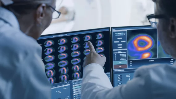
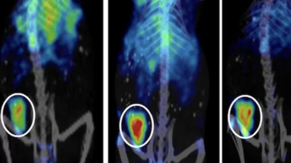
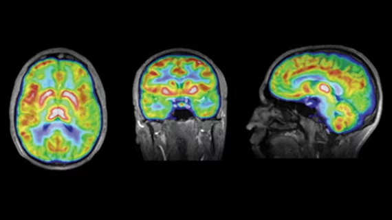
Molecular imaging (also called nuclear medicine or nuclear imaging) can image the function of cells inside the body at the molecular level. This includes the imaging modalities of positron emission computed tomography (PET) and single photon emission computed tomography (SPECT) imaging. How does PET and SPECT imaging work? Small amounts of radioactive material (radiopharmaceuticals) injected into a patient. These can use sugars or chemical traits to bond to specific cells. The radioactive material is taken up by cells that consume the sugars. The radiation emitted from inside the body is detected by photon detectors outside the body. Computers take the data to assemble images of the radiation emissions. Nuclear images may appear fuzzy or ghostly rather than the sharper resolution from MRI and CT.  But, it provides metabolic information at a cellular level, showing if there are defects in the function of the heart, areas of very high metabolic activity associated with cancer cells, or areas of inflammation, data not available from other modalities. These noninvasive imaging exams are used to diagnose cancer, heart disease, Alzheimer’s and Parkinson’s disease, bone disorders and other disorders. 

Thumbnail

Paul Ryan selected to SHINE’s board of directors

In July, SHINE was one of three companies to receive $15 million in funding from the Department of Energy (DOE)’s National Nuclear Security Administration to bolster its production of molybdenum-99 (Mo-99).

Thumbnail

New PET center combines radiology, psychiatry to combat opioid use disorder

The Penn PET Addiction Center of Excellence (PACE) will harness the minds of radiology and psychiatry researchers to better understand opioid use disorder and develop improvement treatment options.

NRC proposes updated guidance for patients receiving radioactive material

The U.S. Nuclear Regulatory Commission (NRC) is asking for comments on its proposed revision to be submitted by Aug. 26.

Thumbnail

FDG-PET beats amyloid imaging for assessing Alzheimer’s

New research suggests that fluorodeoxyglucose (FDG) PET is better for assessing the severity of Alzheimer’s disease and mild cognitive impairment (MCI) than florbetapir-PET scans.

Thumbnail

Should everyone at risk of Alzheimer’s get a brain scan?

A recent story in the New York Times analyzed the positives and potential negatives of undergoing an amyloid PET scan to gain insight into one’s risk of developing Alzheimer’s.

Thumbnail

ASRT backs radiopharmaceutical reform bill

The American Society of Radiologic Technologists (ASRT) voiced their support for recent legislation that seeks to improve patient-access to radiopharmaceuticals in an Aug. 1 statement.

Thumbnail

Novel PET method improves detection of prostate cancer recurrence

Researchers compared prostate-specific membrane antigen (PSMA) PET/CT to traditional 18F-fluciclovine PET/CT for the detection of biochemically recurrent prostate cancer.

Novel imaging method limits radiation dosage from PET, SPECT scans

A team of Japan-based researchers have created a medical camera capable of detecting and imaging radiotracers used in both PET and SPECT scans with limited radiation dosage.