Molecular Imaging

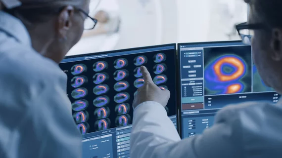
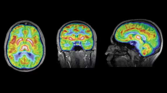
Molecular imaging (also called nuclear medicine or nuclear imaging) can image the function of cells inside the body at the molecular level. This includes the imaging modalities of positron emission computed tomography (PET) and single photon emission computed tomography (SPECT) imaging. How does PET and SPECT imaging work? Small amounts of radioactive material (radiopharmaceuticals) injected into a patient. These can use sugars or chemical traits to bond to specific cells. The radioactive material is taken up by cells that consume the sugars. The radiation emitted from inside the body is detected by photon detectors outside the body. Computers take the data to assemble images of the radiation emissions. Nuclear images may appear fuzzy or ghostly rather than the sharper resolution from MRI and CT.  But, it provides metabolic information at a cellular level, showing if there are defects in the function of the heart, areas of very high metabolic activity associated with cancer cells, or areas of inflammation, data not available from other modalities. These noninvasive imaging exams are used to diagnose cancer, heart disease, Alzheimer’s and Parkinson’s disease, bone disorders and other disorders. 

Thumbnail

NIH awards $20M grant to develop PET radiotracers for diagnosing Parkinson’s

“At the end of five years, we hope to have a radioactive tracer that will be able to detect Parkinson’s early on and provide detailed information about the disease’s progression, which is critical for discovering and testing new treatments," said Robert H. Mach, PhD, a researcher involved in the project.

SHINE closes $50M in financing to support isotope production

The funds, managed by Oaktree Capital Management, will support SHINE’s commercialization of molybdenum-99 and lutetium-177 (Lu-177) in addition to construction of the physical production facility.

Thumbnail

Quantitative differences in PET/CT protocols ‘concerning,’ researchers find

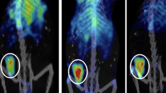
Standardizing protocols for preclinical PET/CT imaging can help translate research findings to the clinical setting, according to a study published Sept. 27 in the Journal of Nuclear Medicine.

Thumbnail

PET/CT more accurate for selecting patients for radionuclide therapy

Metrics based on PET/CT imaging can identify neuroendocrine tumor patients who may benefit from peptide receptor radionuclide therapy (PRRT) better than traditional imaging-based scoring measures, according to a study published in the September issue of The Journal of Nuclear Medicine.

Thumbnail

68Ga-PSMA-11 PET/CT offers value for bone metastases in prostate cancer care

Authors of a new study published Sept. 20 in the Journal of Nuclear Medicine argue that gallium-68 (68Ga) prostate-specific membrane antigen (PSMA)-11 PET/CT imaging can offer additional insight into bone metastases in prostate cancer patients and should be more widely used.

Thumbnail

USP postpones new chapters on safety standards for radiopharmaceuticals

Three chapters published in June by The United States Pharmacopeial Convention (USP) are under appeal and postponed "until further notice."

Thumbnail

FDG-PET/CT radiomic features improves lung cancer care

Integrating 18FDG-PET/CT radiomic tumor and bone marrow features can help predict outcomes in patients with non-small cell lung cancer, according to a Sept. 17 study published in Radiology.

Thumbnail

ANSTO confirms another shutdown at Mo-99 facility

The Australian Nuclear Science and Technology Organization (ANSTO) confirmed it has shut down its Lucas Heights nuclear medicine facility following a “mechanical fault,” The Guardian reported. The shutdown puts an added strain on Australia’s already short supply of molybdenum-99 (Mo-99).