Molecular Imaging

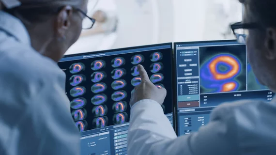
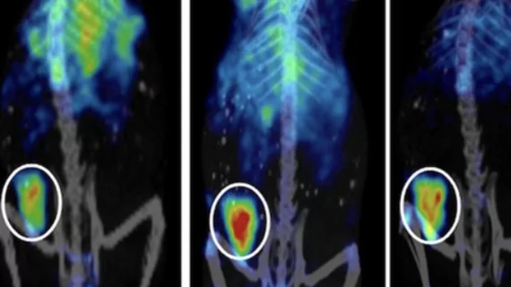
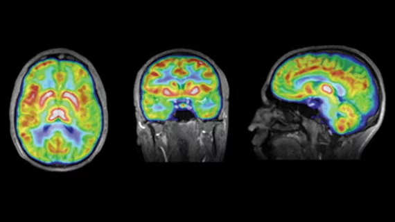
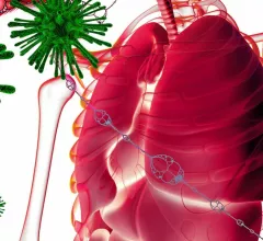
Molecular imaging (also called nuclear medicine or nuclear imaging) can image the function of cells inside the body at the molecular level. This includes the imaging modalities of positron emission computed tomography (PET) and single photon emission computed tomography (SPECT) imaging. How does PET and SPECT imaging work? Small amounts of radioactive material (radiopharmaceuticals) injected into a patient. These can use sugars or chemical traits to bond to specific cells. The radioactive material is taken up by cells that consume the sugars. The radiation emitted from inside the body is detected by photon detectors outside the body. Computers take the data to assemble images of the radiation emissions. Nuclear images may appear fuzzy or ghostly rather than the sharper resolution from MRI and CT.  But, it provides metabolic information at a cellular level, showing if there are defects in the function of the heart, areas of very high metabolic activity associated with cancer cells, or areas of inflammation, data not available from other modalities. These noninvasive imaging exams are used to diagnose cancer, heart disease, Alzheimer’s and Parkinson’s disease, bone disorders and other disorders. 

Radiology efforts over past decade led to 20% drop in patient’s radiation dose, report shows

Radiology has undertaken many efforts to reduce patient exposure to radiation during imaging exams, and findings from a new report suggest those campaigns have made a significant impact.

Thumbnail

Researchers optimize PSMA-targeted prostate cancer therapy to reduce negative side effects

The team found they could maintain the positive impact of therapy, while also minimizing its adverse effects.

Thumbnail

Algorithm reduces radiation dose for molecular breast imaging

The algorithm can also reduce imaging time and improve the patient experience.

Novel PET tracer may enhance lung cancer care

PET using a new voltage-sensitive tracer displayed promise for analyzing activity inside the mitochondria of lung cancer tumors, information that may be valuable for predicting a patient's response to treatment.

Thumbnail

Patient-specific dose measurement improves therapy for neuroendocrine tumors

A team of Swedish researchers found that a hybrid planar and SPECT imaging method fell short in accurately measuring the absorbed treatment dose in some patients, but importantly, performed well in those with bone marrow metastases.

Thumbnail

‘One of the rarest medical isotopes in the world’: Canadian orgs partner to advance cancer research

Canadian Nuclear Laboratories and TRIUMF, the country’s particle accelerator center, have successfully produced actinium-225—a rare isotope that can be used for novel cancer therapy treatments.

Thumbnail

Protective methods to measure radioiodine exposure may be inadequate

Exposure to radioiodine can lead to DNA damage in thyroid cells and other tissue, but new research suggests current radiation protection principles may not be sufficiently monitoring radiation exposure to nuclear medicine professionals.

Thumbnail

PET tracer detects various forms of cancer, lung disease

Results from a study published Oct. 14 in Nature Communications showed the tracer could identify pancreatic, cervical and lung cancer, in addition to a lung tissue disease called idiopathic pulmonary fibrosis.