Molecular Imaging

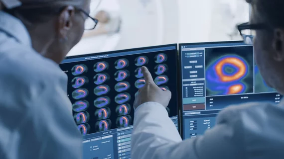
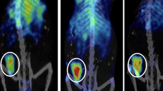
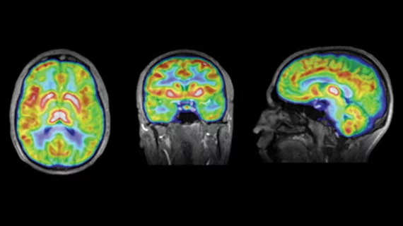
Molecular imaging (also called nuclear medicine or nuclear imaging) can image the function of cells inside the body at the molecular level. This includes the imaging modalities of positron emission computed tomography (PET) and single photon emission computed tomography (SPECT) imaging. How does PET and SPECT imaging work? Small amounts of radioactive material (radiopharmaceuticals) injected into a patient. These can use sugars or chemical traits to bond to specific cells. The radioactive material is taken up by cells that consume the sugars. The radiation emitted from inside the body is detected by photon detectors outside the body. Computers take the data to assemble images of the radiation emissions. Nuclear images may appear fuzzy or ghostly rather than the sharper resolution from MRI and CT.  But, it provides metabolic information at a cellular level, showing if there are defects in the function of the heart, areas of very high metabolic activity associated with cancer cells, or areas of inflammation, data not available from other modalities. These noninvasive imaging exams are used to diagnose cancer, heart disease, Alzheimer’s and Parkinson’s disease, bone disorders and other disorders. 

Thumbnail

NorthStar wraps up construction of new Mo-99 facility

NorthStar Medical Radioisotopes has finished construction of its new molybdenum-99 (Mo-99) processing facility in Beloit, Wisconsin, the company announced on July 16.

Thumbnail

PET tracer can help ID early signs of MS progression

A novel PET radiotracer can help identify changes in the brain’s grey matter that may be indicative of progressive multiple sclerosis (MS), according to a pilot study published in the July issue of Neurology: Neuroimmunology & Neuroinflammation.

Thumbnail

Interim PET scan during prostate cancer therapy tailors treatment

Adding a scan during a patient’s prostate cancer therapy can help personalize treatment, according to research presented at the Society of Nuclear Medicine and Molecular Imaging (SNMMI)’s 2019 Annual Meeting in Anaheim, Calif.

Thumbnail

SNMMI introduces new leadership at annual meeting

The Society of Nuclear Medicine and Molecular Imaging (SNMMI) introduced a host of new officers, including new president Vasken Dilsizian, MD, during its annual meeting in Anaheim, Calif.

Thumbnail

ANSTO nuclear med facility halts production after 2 workers exposed to dangerous radiation dose

The Australian Nuclear Science and Technology Organization (ANSTO)’s new $168 million nuclear medicine facility halted all production after two employees were exposed to an unsafe dose of radiation on Friday, June 21, according to a statement from the organization.

Thumbnail

Radiopharmaceutical safely monitors rheumatoid arthritis

Intravenous (IV) administration of technetium-99m (99mTc) was shown to be a safe, noninvasive way to observe rheumatoid arthritis disease activity, according to a study presented at the 2019 Annual Meeting of the Society of Nuclear Medicine and Molecular Imaging (SNMMI) in Anaheim, Calif.

Thumbnail

Researchers create new method for developing PET radiotracers

University of North Carolina-Chapel Hill researchers have developed a new method for creating radiotracers used in PET imaging. The technique may improve imaging of diseases such as cancer, according to the study published in Science.

Thumbnail

Novel PET scan improves diagnosis, monitoring of inflammatory bowel disease

Immuno-PET offers a more efficient diagnosis of inflammation in inflammatory bowel disease (IBD) compared to traditional invasive methods, according to a new study published in the June issue of the Journal of Nuclear Medicine.