Molecular Imaging

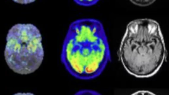
Molecular imaging (also called nuclear medicine or nuclear imaging) can image the function of cells inside the body at the molecular level. This includes the imaging modalities of positron emission computed tomography (PET) and single photon emission computed tomography (SPECT) imaging. How does PET and SPECT imaging work? Small amounts of radioactive material (radiopharmaceuticals) injected into a patient. These can use sugars or chemical traits to bond to specific cells. The radioactive material is taken up by cells that consume the sugars. The radiation emitted from inside the body is detected by photon detectors outside the body. Computers take the data to assemble images of the radiation emissions. Nuclear images may appear fuzzy or ghostly rather than the sharper resolution from MRI and CT.  But, it provides metabolic information at a cellular level, showing if there are defects in the function of the heart, areas of very high metabolic activity associated with cancer cells, or areas of inflammation, data not available from other modalities. These noninvasive imaging exams are used to diagnose cancer, heart disease, Alzheimer’s and Parkinson’s disease, bone disorders and other disorders. 

Thumbnail

Molecular imaging research could find less toxic, more effective breast cancer treatments

Researchers from the Medical University of South Carolina (MUSC) Hollings Cancer Center in Charleston have received a three-year, $1.2 million grant from the U.S. Department of Defense to use molecular imaging tools to test antibody therapies for breast cancer—potentially producing fewer side effects.

Thumbnail

NorthStar may receive federal funding to produce Mo-99

NorthStar Medical Radioisotopes in Beloit, Wis. and the Janesville, Wis.-planned SHINE Medical Technologies may receive a portion of the $20 million in federal technology funding bill approved by President Donald Trump to produce molybdenum-99 (Mo-99).

Thumbnail

Nanoplatform developed with 3 molecular imaging modalities may improve cancer diagnosis

A new hybrid nanoplatform that simultaneously uses three types of imaging modalities—MRI, CT and fluorescence optical imaging—to locate tumors could give a new edge to molecular imaging and tumor diagnosis.  

Thumbnail

Cancer imaging researcher receives highest honor from World Molecular Imaging Society

The World Molecular Imaging Society (WMIS) has awarded Robert Gillies, PhD, the 2018 Gold Medal Award to honor his lifetime of research in cancer molecular imaging and cancer biology, according to a WMIS press release.

Thumbnail

Imaging blood-brain barrier as biomarker could ID earliest warning signs of Alzheimer’s

Alzheimer’s could be diagnosed earlier if the blood-brain barrier was monitored as a biomarker and potential drug target using MRI or PET, according to research published online Sept. 24 in Nature Neuroscience.

Thumbnail

BlueCross BlueShield Tennessee to expand cardiac PET coverage

BlueCross BlueShield of Tennessee announced it will expand Medicare coverage for cardiac PET, according to the Society for Nuclear Medicine and Molecular Imaging (SNMMI). The change will take effect later this year.

Thumbnail

Tau PET agent accurately diagnosis Alzheimer’s from other neurodegenerative diseases

In patients with diagnosed Alzheimer’s disease (AD), [18F] flortaucipir PET imaging proved accurate in distinguishing AD from other neurodegenerative diseases, according to a multicenter study published in JAMA.

SNMMI, ASNC encourage BCBS to expand coverage of cardiac PET

The Society of Nuclear Medicine and Molecular Imaging (SNMMI) and the American Society of Nuclear Cardiology (ASNC) submitted a letter to BlueCross BlueShield (BCBS) of Tennessee urging an expansion of cardiac PET coverage.