Molecular Imaging

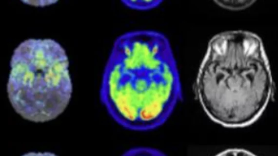
Molecular imaging (also called nuclear medicine or nuclear imaging) can image the function of cells inside the body at the molecular level. This includes the imaging modalities of positron emission computed tomography (PET) and single photon emission computed tomography (SPECT) imaging. How does PET and SPECT imaging work? Small amounts of radioactive material (radiopharmaceuticals) injected into a patient. These can use sugars or chemical traits to bond to specific cells. The radioactive material is taken up by cells that consume the sugars. The radiation emitted from inside the body is detected by photon detectors outside the body. Computers take the data to assemble images of the radiation emissions. Nuclear images may appear fuzzy or ghostly rather than the sharper resolution from MRI and CT.  But, it provides metabolic information at a cellular level, showing if there are defects in the function of the heart, areas of very high metabolic activity associated with cancer cells, or areas of inflammation, data not available from other modalities. These noninvasive imaging exams are used to diagnose cancer, heart disease, Alzheimer’s and Parkinson’s disease, bone disorders and other disorders. 

CMS denies coverage for NaF PET exams leaving experts, imaging groups confused

In a blow to Medicare patients with metastatic cancers, the Centers for Medicare & Medicaid Services (CMS) announced it has rejected reimbursement coverage for 18F-sodium fluoride positron emission tomography (NaF PET) imaging.

Thumbnail

UVA researchers release guidelines for x-ray crystallography, cancer research

New guidelines for researchers wanting to study how molecules in the body use metals such as iron and magnesium to maintain good health have been established by researchers at the University of Virginia (UVA).

Radiopharmaceutical can ease symptoms in some cancers, improve survival outlook

A team of scientists found therapeutic treatment using radiopharmaceutical I-131 mIBG can effectively mitigate symptoms of certain neuroendocrine tumors and resulted in improved prognosis for patients with symptomatic progression, radiographic response or stability and biochemical response.

Thumbnail

Diamonds may be a sparkling method to reduce costs of medical imaging, drug studies

A new discovery involving diamonds may significantly cut costs related to medical imaging and drug-discovery devices, according to a team of researchers led by the U.S. Department of Energy and the University of California, Berkeley. 

Researchers create novel method predicting tumor response in radioligand therapy

A team of German researchers used PET/CT and modeling practices to create a new method to pattern and predict tumor response in radioligand therapy (RLT), which authors say is the first attempt to do so.

Thumbnail

Could a breast imaging pill eliminate the need for annual mammograms?

The "disease screening pill," developed by researchers from the University of Michigan, lit up malignant tumors in mice when exposed to near-infrared light—all without the use of radiation, according to a study published in Molecular Pharmaceutics.

Thumbnail

SNMMI collaborates with industry leaders to advance molecular imaging

On May 17, the Society of Nuclear Medicine and Molecular Imaging (SNMMI) announced the Value Initiative Industry Alliance—a collaboration among corporate community members that will help implement the organization’s Value Initiative and advance nuclear medicine.

Thumbnail

PET tracer may predict response to therapy in bone-dominant breast cancer patients

A prospective comparison study of two PET tracers found 18F-FDG could provide valuable measures of activity in bone-dominant (BD) metastatic breast cancer (MBC) patients, while also predicting responses to therapy.