Molecular Imaging

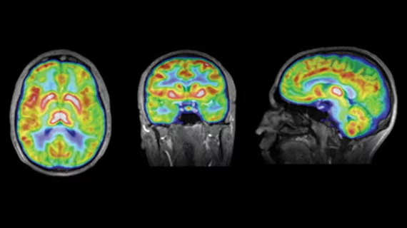
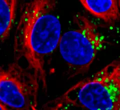
Molecular imaging (also called nuclear medicine or nuclear imaging) can image the function of cells inside the body at the molecular level. This includes the imaging modalities of positron emission computed tomography (PET) and single photon emission computed tomography (SPECT) imaging. How does PET and SPECT imaging work? Small amounts of radioactive material (radiopharmaceuticals) injected into a patient. These can use sugars or chemical traits to bond to specific cells. The radioactive material is taken up by cells that consume the sugars. The radiation emitted from inside the body is detected by photon detectors outside the body. Computers take the data to assemble images of the radiation emissions. Nuclear images may appear fuzzy or ghostly rather than the sharper resolution from MRI and CT.  But, it provides metabolic information at a cellular level, showing if there are defects in the function of the heart, areas of very high metabolic activity associated with cancer cells, or areas of inflammation, data not available from other modalities. These noninvasive imaging exams are used to diagnose cancer, heart disease, Alzheimer’s and Parkinson’s disease, bone disorders and other disorders. 

Thumbnail

Molecular imaging becomes 3D interactive experience with VR

A team of researchers has created a virtual reality (VR) environment that allows users to manipulate 3D molecular proteins 10 times faster than with a 2D screen, according to a July 3 article by the New York Times.

Thumbnail

Fluciclovine PET/CT IDs recurrent prostate cancer, alters patient management

Adding fluorine-18 (18F)-fluciclovine PET/CT to the diagnostic treatment plan of patients with biochemical recurrence of prostate cancer can identify previously undetected lesions and alter treatment management for patients with the disease.

Thumbnail

MR-assisted PET data optimization may improve neuroanalysis of dementia patients

Researchers—led by Kevin Chen, PhD, a postdoctoral fellow at Harvard University's Microrobotics Lab—have found that spatiotemporally correlated data acquired using a single magnetic resonance (MR) sequence may be successfully used in a PET/MRI scanner for PET attenuation, motion and partial effects corrections.

Thumbnail

Satoshi Minoshima named president of SNMMI

A new group of officers has been selected at the Society of Nuclear Medicine and Molecular Imaging (SNMMI) 2018 annual meeting, including president Satoshi Minoshima, MD, PhD, professor and chair of the department of radiology and imaging sciences at the University of Utah in Salt Lake City.

Novel PET, SPECT techniques help track, modify T cells for immunotherapy

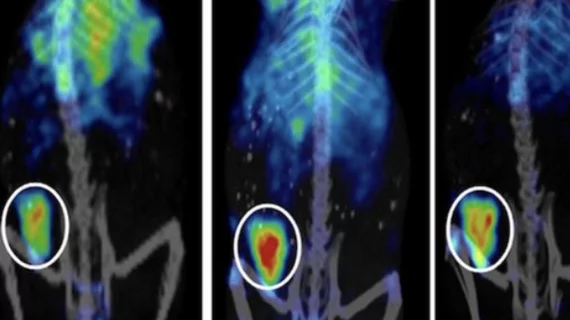
With PET and single-photon emission computed tomography (SPECT), researchers have demonstrated that T cells can be modified with the antibody report 1 (DAbR1) gene to enable in vivo tracking for immunotherapy, according to research published in the Journal of Nuclear Medicine.

At SNMMI 2018, Siemens Healthineers Announces FDA Clearance of New Molecular Imaging Functionalities

At the 2018 Annual Meeting of the Society of Nuclear Medicine and Molecular Imaging (SNMMI), June 23-26 at the Pennsylvania Convention Center in Philadelphia, Siemens Healthineers announces the Food and Drug Administration (FDA) clearance of four new system features for the Biograph mCT family of positron emission tomography/computed tomography (PET/CT) systems

Rise of PET: 3 areas where nuclear imaging can boost patient-care

Positron emission tomography (PET) has historically been an afterthought in musculoskeletal imaging for many logical reasons, but authors of a new investigation believe the method should play a larger role in managing these patients.

Thumbnail

SNMMI backs updated ASNC guidelines on SPECT MPI

New 2018 guidelines for myocardial perfusion imaging (MPI) protocols on conventional and novel single photon emission computed tomography (SPECT) for nuclear cardiology practitioners has received praise from the Society for Nuclear Medicine and Molecular Imaging (SNMMI).