Molecular Imaging

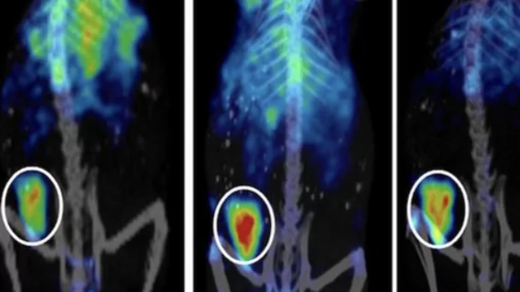
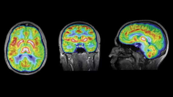
Molecular imaging (also called nuclear medicine or nuclear imaging) can image the function of cells inside the body at the molecular level. This includes the imaging modalities of positron emission computed tomography (PET) and single photon emission computed tomography (SPECT) imaging. How does PET and SPECT imaging work? Small amounts of radioactive material (radiopharmaceuticals) injected into a patient. These can use sugars or chemical traits to bond to specific cells. The radioactive material is taken up by cells that consume the sugars. The radiation emitted from inside the body is detected by photon detectors outside the body. Computers take the data to assemble images of the radiation emissions. Nuclear images may appear fuzzy or ghostly rather than the sharper resolution from MRI and CT.  But, it provides metabolic information at a cellular level, showing if there are defects in the function of the heart, areas of very high metabolic activity associated with cancer cells, or areas of inflammation, data not available from other modalities. These noninvasive imaging exams are used to diagnose cancer, heart disease, Alzheimer’s and Parkinson’s disease, bone disorders and other disorders. 

UltraSPECT Installs 450th System in USA At Firelands Regional Medical Center

UltraSPECT partners with HeartLight Pharmacy Services to help nuclear medicine facilities deliver reduced radiation dose, shortened scan time for patient, staff safety.

Thumbnail

The University of California – San Francisco to Install MILabs VECTor4CT in Support of Expanding Molecular and Functional Imaging Research

The Center for Molecular and Functional Imaging (CMFI) at the University of California – San Francisco will install the MILabs VECTor4CT in support of a large number of NIH funded research projects in the areas of oncology, neuroscience and drug discovery/development.  The UCSF Department of Radiology and Biomedical Imaging invested significant resources in establishing the CMFI in 2003.  Major research efforts at CMFI include translational nuclear medicine imaging research using both SPECT/CT and PET/CT.  Utilization of the VECTor4CT will benefit from the CMFI installed medical cyclotron, state of the art radiopharmaceutical laboratory and in-house vivarium all under one roof.

Thumbnail

Annual International Summit “Ageing” to be Held from February 9 to 11, 2016

Ageing 2016 will be held from February 9 to 11, 2016 at Cineworld: The O2 Peninsula Square London SE10 0DX United Kingdom. The annual international summit will look at the various aspects of processes and research involved in ageing and senescence.

Thumbnail

Study indicates mentally demanding tasks can protect brain power

Mentally demanding activities may be neuroprotective and an important element for maintaining a healthy brain into late adulthood, according to findings published in Restorative Neurology and Neuroscience.

Thumbnail

Cancer surveillance PET rises, suggesting overutilization

A robust, population-based study has revealed that, from 2001 through 2009, the use of PET imaging swelled among patients with resected non–small-cell lung cancer (NSCLC) and colorectal cancer (CRC).

Study: Gadolinium has little impact on accuracy of MRI for pediatric tumors

Gadolinium chelate administration is largely unnecessary for imaging pediatric tumors when performing MRI or combined PET/MR scans, according to a recent study published by the Journal of Nuclear Medicine.

Thumbnail

A ‘nuclear scalpel’ may emerge from discovery of four new elements

Scientists from Japan, Russia and the U.S. have discovered four new chemical elements that, taken together, complete the seventh period of the periodic table of elements. And the first area of human endeavor to find a practical use for the expanded knowledge may be nuclear medicine.

Adding whole-body PET/MRI improves evaluation of colorectal cancer

Compared with conventional multi-detector, contrast-enhanced CT (CECT) alone, CECT with integrated whole-body PET/MRI is better at detecting metastatic lesions and characterizing indeterminate lesions in the colon.