Molecular Imaging

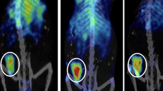
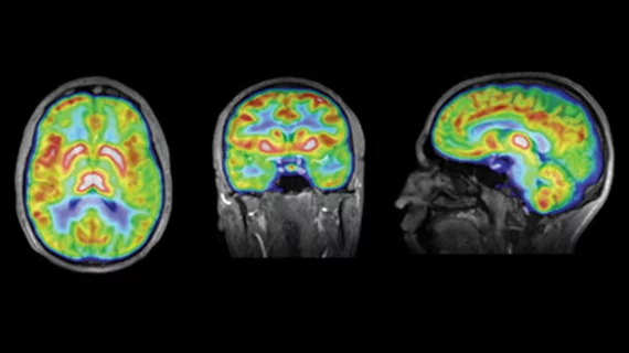
Molecular imaging (also called nuclear medicine or nuclear imaging) can image the function of cells inside the body at the molecular level. This includes the imaging modalities of positron emission computed tomography (PET) and single photon emission computed tomography (SPECT) imaging. How does PET and SPECT imaging work? Small amounts of radioactive material (radiopharmaceuticals) injected into a patient. These can use sugars or chemical traits to bond to specific cells. The radioactive material is taken up by cells that consume the sugars. The radiation emitted from inside the body is detected by photon detectors outside the body. Computers take the data to assemble images of the radiation emissions. Nuclear images may appear fuzzy or ghostly rather than the sharper resolution from MRI and CT.  But, it provides metabolic information at a cellular level, showing if there are defects in the function of the heart, areas of very high metabolic activity associated with cancer cells, or areas of inflammation, data not available from other modalities. These noninvasive imaging exams are used to diagnose cancer, heart disease, Alzheimer’s and Parkinson’s disease, bone disorders and other disorders. 

HTG Molecular Diagnostics Announces VERI/O Laboratory Service

TUCSON, Ariz., June 23, 2016 (GLOBE NEWSWIRE) -- HTG Molecular Diagnostics, Inc. (Nasdaq:HTGM), a provider of instruments and reagents for molecular profiling applications, today announced the introduction of its Tucson‑based VERI/O laboratory service, offering laboratory support for its biopharma clients in biomarker research and companion diagnostic development.  The VERI/O laboratory service formalizes and expands HTG’s traditional service offerings, including molecular profiling of retrospective cohorts to support development of targeted and immuno-oncology therapies, building custom research-use-only (RUO) assays to support early stage clinical programs, and developing companion diagnostic assays for use in Phase III registration trials.

MR Solutions offers choice of two preclinical cryogen-free MRI scanner ranges

MR Solutions, which introduced cryogen-free MRI scanners in 2012, is now offering a choice of two cryogen-free preclinical scanner ranges – the Flexiscan and Powerscan. The Flexiscan range is available with multi-modality accessories which can be mixed and matched according to research requirements. It is a flexible MRI system as it can incorporate integrated multi-modality options such as PET or SPECT. The Flexiscan system requires no specialist knowledge and can be operated simply by running pre-defined settings. There is no need to materially alter the parameters of the machine.

Study: Single season of high school football can damage athlete's brain

The repeated impacts experienced in one season of playing high school football can do significant damage to an athlete’s brain, according to a recent study published by the Journal of Neurotrauma.

Molecular Devices Launches High-Content Imaging System with New 3D Analysis Software

Sunnyvale, Calif. – April 19, 2016 – Molecular Devices, LLC, a leader in protein and cell biology technologies, today announced the launch of its ImageXpress® Micro 4 High-Content Imaging System and revolutionary 3D image analysis capability in the latest version of MetaXpress® High-Content Image Acquisition and Analysis Software. For scientists conducting basic research or high-throughput screening, the ImageXpress System is a premiere, integrated and scalable toolset for optimal 3D image acquisition and analysis.

ABT Partners to Establish the First Radiopharmaceutical Production Facility in Cyprus

Knoxville, TN, UNITED STATES, March 18, 2016– ABT Molecular Imaging has partnered with a world-renowned oncology group to install the BG-75 Biomarker Generator System in  The Republic of Cyprus.  The BG-75 integrates a compact cyclotron, micro-chemistry, and automated quality control, to provide on-demand F-18 FDG production in one seamless solution to support the implementation of PET/CT.

New fluorine probe called a ‘major leap’ for MRI sensitivity

Radiology and biochemistry researchers at UC-San Diego say they have figured out how to dissolve and encapsulate metals inside fluorine-based droplets, in the process synthesizing a new cell-labeling probe that can, in in effect, “greatly amp up the MRI signal” for potential use in in immunotherapy, inflammation treatment and other emerging clinical applications. 

Thumbnail

Your full DNA is in your smartphone, or can be

Want to know which specific hereditary ailments you, and you alone, are susceptible to? All of them? 

Study: New peptide probes could help detect disease faster

A new advancement in real-time visualization of body tissues using molecular imaging probes could significantly increase early detection and treatment capabilities of cancer and other diseases, according to results of a study published online in Nature Communications.