Molecular Imaging

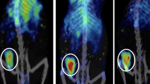
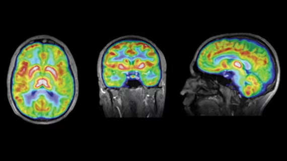
Molecular imaging (also called nuclear medicine or nuclear imaging) can image the function of cells inside the body at the molecular level. This includes the imaging modalities of positron emission computed tomography (PET) and single photon emission computed tomography (SPECT) imaging. How does PET and SPECT imaging work? Small amounts of radioactive material (radiopharmaceuticals) injected into a patient. These can use sugars or chemical traits to bond to specific cells. The radioactive material is taken up by cells that consume the sugars. The radiation emitted from inside the body is detected by photon detectors outside the body. Computers take the data to assemble images of the radiation emissions. Nuclear images may appear fuzzy or ghostly rather than the sharper resolution from MRI and CT.  But, it provides metabolic information at a cellular level, showing if there are defects in the function of the heart, areas of very high metabolic activity associated with cancer cells, or areas of inflammation, data not available from other modalities. These noninvasive imaging exams are used to diagnose cancer, heart disease, Alzheimer’s and Parkinson’s disease, bone disorders and other disorders. 

Thumbnail

New FDA draft guidance covers IDE applications for neurological medical devices

The FDA has issued a new draft guidance intended to assist FDA staff and those in the healthcare industry with submitting investigational device exemption (IDE) applications to conduct clinical trials for neurological medical devices. 

Thumbnail

Twitter for clinical-trial recruitment: All but untapped, barely even explored

Of more than 1,500 tweets randomly pulled from a qualified starting cohort of more than 15,000 mentioning the term “lung cancer” over two and a half weeks in January 2015, just one lonely tweet linked to a patient recruitment website for a clinical trial.

Predicting liver tumor recurrence with immediate post-ablation FDG PET/CT

Performing PET/CT examinations using the radiotracer 18-Fluoro-deoxyglucose (FDG) immediately following localized liver tumor ablation is more effective than contrast-enhanced CT for predicting tumor recurrence, according to results of a study published online in the Journal of Nuclear Medicine.

Consultant to concerned residents: N.J. radiopharmaceutical facility not a threat

Patrick McDermott, a Rutgers University scientist who specializes in ionized radiation, has said a radiopharmaceutical factory scheduled to open soon in Millburn, N.J., poses no health risks to the area. McDermott was hired back in October 2015 to assess the safety of the factory and reported his findings this week.

Thumbnail

‘Mini-brains’ spare the research animals, spread the neuroscience

Johns Hopkins researchers have begun genetically modifying adult cells to create balls of neurons that may serve as “mini-brains,” potentially good for preclinically studying—in petri dishes rather than in animals—Alzheimer’s, Parkinson’s, autism and the effects of drugs, for starters.

Thumbnail

Amyloid PET with automated analysis excels at ruling out Alzheimer’s

When it comes to predicting the cognitive-impairment course Alzheimer’s disease will take in a mildly stricken patient, neither of two tracers commonly used with PET—18F-FDG (for assessing glucose metabolism) and 11C-PiB (for assessing amyloid deposits)—brings much to the table. And that’s so regardless of automated vs. visual analysis.

Thumbnail

Siemens Healthcare Limited launches new assay range to improve workload consolidation in the laboratory

A range of new assays has been released by Siemens for use on ADVIA Centaur and IMMULITE XPi 2000 systems.Launch includes the first automated quantitative thyroid stimulating immunoglobulin (TSI) assay used in the diagnosis of Graves’ disease.

Thumbnail

Brain injuries may remain unhealed long enough to increase Alzheimer’s risk

Preliminary research at Imperial College London has shown that increased levels in binding of the Alzheimer’s-associated radioligand 11C-PiB—the go-to radioactive biochemical substance in PET imaging for cerebral amyloid beta plaque—also show up in people who have had traumatic brain injury, and for more than a decade after the TBI event.