Magnetic Resonance Imaging

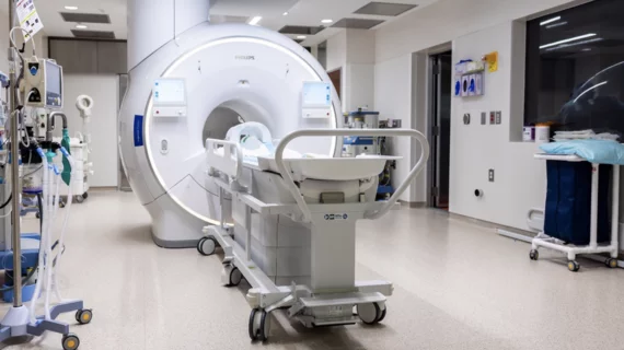
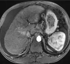
Magnetic resonance imaging (MRI) is the gold standard imaging modality for soft tissues. It produces detail cross-sectional images of soft tissue and bone anatomy, including muscles, tendons, ligaments, brain and organs, without the use of ionizing radiation. In addition to orthopedic imaging, MRI is also used for heart, brain and breast. MRI uses gadolinium contrast in many exams to highlight tissues and blood vessels, which enhances images and offers better diagnostic quality. It can also be used in conjunction with PET scans. How does MRI work? MR creates images by using powerful magnets to polarize hydrogen atoms in water (the body is made of of more than 80% water) so they face in one direction. A radiofrequency pulse is then used to ping these atoms, causing them to wobble, or resonate. The MRI coils detect this and computers can assemble images from the signals. Basic MRI scans will focus on the resonance of fat and water in two different sequences, which highlight and contrast different features in the anatomy.

COVID-19 vaccine associated myocarditis on short-axis 1.5T MRI images of a 19-year-old man who presented with chest pain three days following the second dose of an mRNA COVID-19 vaccine.

New cardiac MRI analysis offers updated insight into long-term impact of vaccine-related myocarditis

Months after their initial myocarditis diagnosis, late gadolinium enhancement (LGE) was observed in 62% of patients on follow-up cardiac MRI.

Foot ultrasound bested foot MRI on pooled sensitivity while lagging on specificity

MRI beats ultrasound at grading common foot injury, but tables turn for up-or-down diagnostics

Injuries of the plantar plate—the cluster of ligaments underneath the four smaller “knuckles” of the toes—are better assessed with MRI than with ultrasound scans. 

Thumbnail

Cardiac implantable electronic devices can be safely reprogrammed to MRI-safe mode via remote operator

In 209 study participants, the remote reprogramming resulted in a successful, event-free MRI scan for every patient.

Thumbnail

AI looking handy with 3D abdominal ultrasound

Mayo Clinic researchers have demonstrated a deep learning model that can automatically segment kidneys and measure total kidney volumes using only 3D ultrasound images.

Thumbnail

Abbreviated breast MRI deemed an attractive screening option—sometimes

AB-MRI is a cost-effective means of screening women with dense breast tissue for breast cancer—as long as the per-exam costs don’t top 82% of what would have been spent to perform full-protocol breast MRI.

Ischemic stroke CT imaging. Images courtesy of RSNA

Stroke care still lags among certain Medicare populations

Despite increases of 250% for CTA and 428% for CTP, researchers observed lower rates of neuroimaging utilization among rural residents, older individuals (80 years and above), women and Black patients.

Thumbnail

Memory complaints associated with structural brain abnormalities and increased dementia risk

In participants who reported concern about their subjective memory, higher white matter hyperintensity volumes were observed and those individuals exhibited 174% faster cognitive decline than their peers.

Study links MRI findings with mental health disorders

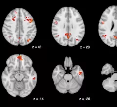
Based on imaging, researchers suggested that gray matter volume could be a predictive marker of outcomes in mental health disorders, such as depression.