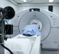
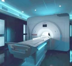
Magnetic Resonance Imaging

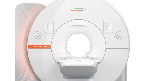
Magnetic resonance imaging (MRI) is the gold standard imaging modality for soft tissues. It produces detail cross-sectional images of soft tissue and bone anatomy, including muscles, tendons, ligaments, brain and organs, without the use of ionizing radiation. In addition to orthopedic imaging, MRI is also used for heart, brain and breast. MRI uses gadolinium contrast in many exams to highlight tissues and blood vessels, which enhances images and offers better diagnostic quality. It can also be used in conjunction with PET scans. How does MRI work? MR creates images by using powerful magnets to polarize hydrogen atoms in water (the body is made of of more than 80% water) so they face in one direction. A radiofrequency pulse is then used to ping these atoms, causing them to wobble, or resonate. The MRI coils detect this and computers can assemble images from the signals. Basic MRI scans will focus on the resonance of fat and water in two different sequences, which highlight and contrast different features in the anatomy.

Radiologists call for standardized imaging criteria for diagnosing pancreatitis in children

A recent analysis conducted on interobserver agreement when diagnosing chronic pediatric pancreatitis has raised some concerns about the need for more standardized diagnostic criteria.

Thumbnail

Understanding the structural factors that fuel racial disparities in the use of prostate MRI

Neighborhood-level socioeconomic status, racialized residential segregation, and socioeconomic status all play a part, according to research published in JAMA Oncology

liver cancer

Patients prioritize higher imaging sensitivity over lower costs when weighing HCC screening options

These results have important implications for understanding patient preferences as determinants of adherence, imaging experts wrote recently. 

Thumbnail

MRIs show how even modest alcohol consumption can impact the brain

Experts analyzed more than 36,000 brain MRIs to determine how modest alcohol intake impacts brain volume and aging.

Thumbnail

Experts unveil NS-RADS, a new MRI-powered system for classifying neuropathy

The Neuropathy Score Reporting and Data System (NS-RADS) was validated by radiologists from varying experience levels with good interrater agreement, experts reported in the American Journal of Roentgenology.

Thumbnail

MRI features uncover differences between the brains of autistic girls and boys

Scientists believe their findings could inform the development of gender-specific diagnosis and treatment strategies for the disorder, according to research published the British Journal of Psychiatry.

'Pandemic brain': PET/MRI images reveal how COVID's impact is felt by non-infected individuals

A new analysis in Brain, Behavior, and Immunity shows that no one is immune to the effects of the ongoing pandemic. 

Deep learning model triages brain MRIs for abnormalities to prioritize reads

The deep learning model was trained to recognize abnormalities in real-time, reducing delays in image interpretation for clinically relevant findings.