Magnetic Resonance Imaging

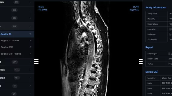
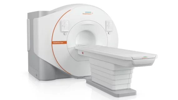
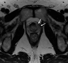
Magnetic resonance imaging (MRI) is the gold standard imaging modality for soft tissues. It produces detail cross-sectional images of soft tissue and bone anatomy, including muscles, tendons, ligaments, brain and organs, without the use of ionizing radiation. In addition to orthopedic imaging, MRI is also used for heart, brain and breast. MRI uses gadolinium contrast in many exams to highlight tissues and blood vessels, which enhances images and offers better diagnostic quality. It can also be used in conjunction with PET scans. How does MRI work? MR creates images by using powerful magnets to polarize hydrogen atoms in water (the body is made of of more than 80% water) so they face in one direction. A radiofrequency pulse is then used to ping these atoms, causing them to wobble, or resonate. The MRI coils detect this and computers can assemble images from the signals. Basic MRI scans will focus on the resonance of fat and water in two different sequences, which highlight and contrast different features in the anatomy.

Thumbnail

Memory complaints associated with structural brain abnormalities and increased dementia risk

In participants who reported concern about their subjective memory, higher white matter hyperintensity volumes were observed and those individuals exhibited 174% faster cognitive decline than their peers.

Study links MRI findings with mental health disorders

Based on imaging, researchers suggested that gray matter volume could be a predictive marker of outcomes in mental health disorders, such as depression.

Thumbnail

New mobile low-field MRI scanner inches closer to increasing imaging access in rural communities

It takes only five minutes for the portable MRI system, "Scan-a-van," to start scanning after arriving at patients' homes.

Thumbnail

Use of unnecessary neuroimaging for patients with dizziness prevalent in outpatient settings

Quality improvement efforts have sought to address such low-value diagnostic testing in the ED, with little attention paid to ambulatory settings, experts wrote in JAMA

Thumbnail

MRI/PET scans link brainstem atrophy to dementia symptoms

Experts suggest that their findings could help differentiate between dementia and other neurological diseases that have similar symptoms.

Thumbnail

Researchers use MRI scans to develop a growth chart specific to the human brain

A team of researchers have developed a new tool that benchmarks brain development and growth based on over 120,000 MRI scans.

Thumbnail

Even low levels of prenatal alcohol exposure affect brain development, MRI scans show

Researchers revealed the MRI findings were associated with worse externalizing behavior scores in children with prenatal alcohol exposure and are a cause of concern for pediatric brain development.

Thumbnail

Vaping and smoking have opposite physiologic effects on the lungs, MRI scans show

Researchers found that tobacco exposure decreased lung perfusion, while vaping led to an increase after just one smoking session.