Magnetic Resonance Imaging

Magnetic resonance imaging (MRI) is the gold standard imaging modality for soft tissues. It produces detail cross-sectional images of soft tissue and bone anatomy, including muscles, tendons, ligaments, brain and organs, without the use of ionizing radiation. In addition to orthopedic imaging, MRI is also used for heart, brain and breast. MRI uses gadolinium contrast in many exams to highlight tissues and blood vessels, which enhances images and offers better diagnostic quality. It can also be used in conjunction with PET scans. How does MRI work? MR creates images by using powerful magnets to polarize hydrogen atoms in water (the body is made of of more than 80% water) so they face in one direction. A radiofrequency pulse is then used to ping these atoms, causing them to wobble, or resonate. The MRI coils detect this and computers can assemble images from the signals. Basic MRI scans will focus on the resonance of fat and water in two different sequences, which highlight and contrast different features in the anatomy.

Thumbnail

New MRI technique 'lights up' prostate cancer

Scientists involved in the study believe the innovative technology has exciting potential to improve screening, prognosis and treatment, according to research published in Scientific Reports. 

quality

Study charts uptick in unnecessary CT, MR imaging surveillance of noninvasive bladder cancer

The impact of these patterns is substantial and may have negative consequences for patients and the healthcare system, experts wrote in JAMA Network Open

Thumbnail

Is MRI a suitable alternative to CT for testicular cancer surveillance? Research offers insight

CT surveillance is the standard of care for postoperatively monitoring testicular cancer, but when patients must undergo scans every few months after surgery, accumulative radiation exposure becomes a concern.

Size matters when it comes to neuroimaging studies

New research published in Nature indicates that the results of many neuroimaging studies lack reliability due to their inadequate sample sizes.

How second opinions from subspecialty radiologists alter cancer care

Such reports sometimes unearth widespread variability in in the quality of outside imaging exams, a significant amount of which result in treatment changes.

A dedicated Cardiac MRI scanner. The presence of myocardial fibrosis on cardiac MRI can help anticipate adverse arrhythmic events and  may offer a way to improve patient selection for cardiac implantable electronic devices (CIEDs). This is a dedicated cardiac MRI system at Baylor Scott White.

AI tool detects CVD on cardiac MRI in 20 seconds with high precision

The algorithm is already in use at some institutions in Europe, with plans for it to be utilized globally at many more within the year.

Multiple sclerosis ribbon MS

Radiologist diagnoses multiple sclerosis via MRI, but patient doesn’t find out until 8 years later

A Massachusetts multispecialty center has dodged a malpractice suit after the patient missed the state's seven-year deadline to file. 

Thumbnail

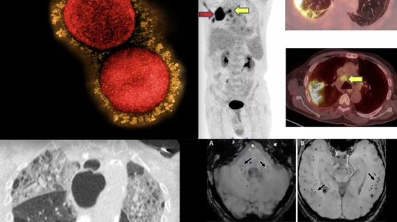
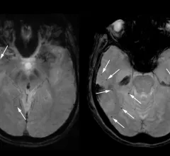
MRI scans show COVID's 'significant' impact on the brain

Even individuals who had recovered from what would be considered a mild case of COVID displayed signs of tissue damage and reductions in gray matter and overall brain size, experts reported in Nature.