Magnetic Resonance Imaging

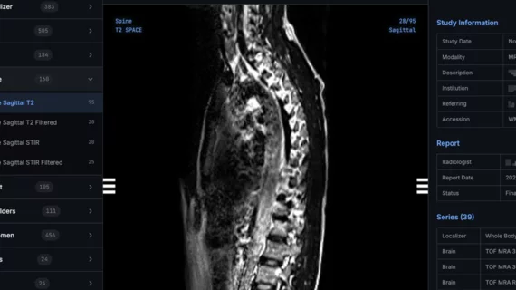
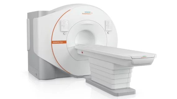
Magnetic resonance imaging (MRI) is the gold standard imaging modality for soft tissues. It produces detail cross-sectional images of soft tissue and bone anatomy, including muscles, tendons, ligaments, brain and organs, without the use of ionizing radiation. In addition to orthopedic imaging, MRI is also used for heart, brain and breast. MRI uses gadolinium contrast in many exams to highlight tissues and blood vessels, which enhances images and offers better diagnostic quality. It can also be used in conjunction with PET scans. How does MRI work? MR creates images by using powerful magnets to polarize hydrogen atoms in water (the body is made of of more than 80% water) so they face in one direction. A radiofrequency pulse is then used to ping these atoms, causing them to wobble, or resonate. The MRI coils detect this and computers can assemble images from the signals. Basic MRI scans will focus on the resonance of fat and water in two different sequences, which highlight and contrast different features in the anatomy.

MRI findings associated with poor thrombectomy outcomes after stroke

For patients who have suffered an acute ischemic stroke, white matter hyperintensity visualized on MRI before thrombectomy might be indicative of poor post-procedure clinical outcomes.

Thumbnail

Augmented reality bear helps calm pediatric patients undergoing MRI exams

Choco the bear appears before children in real time via a tablet to explain the MRI process and even completes an exam himself.

Thumbnail

Water-fat separation sequence yields superior image quality compared to standard coronary MRA

The 3-T Dixon GRE magnetic resonance angiography method produced better image quality and yielded greater overall diagnostic performance, according to a new study in AJR.

Thumbnail

MRI scans link atypical growth of key brain structure during infancy with autism

Experts noted that this overgrowth occurs before the onset of behavioral symptoms associated with autism and can be identified in babies between 6 and 12 months of age.

quality imaging appropriateness clinical decision support CAS AUC

Application for reporting adverse contrast reactions significantly increases radiologists’ thoroughness

Mass General has piloted the use of a new tool called CISaR (Contrast Incident Support and Reporting) to vastly improve documentation.

button battery

MRI after button battery removal: Consensus on next steps lacking among referrers

There are no published guidelines on how imaging should inform clinical management, Seattle Children's Hospital experts noted. 

Thumbnail

Misuse of public imaging data is producing 'overly optimistic' results in machine learning research

 "This research aims to raise a red flag regarding naive off-label usage of open-access data in the development of machine-learning algorithms," experts involved in the study said.

Thumbnail

Diffusion tensor imaging of the knee predicts pediatric bone growth

Current bone age-based methods of growth prediction in children are inaccurate and frequently overestimate final height, experts explained in Radiology.