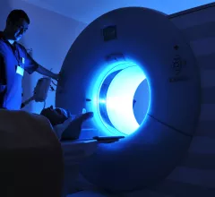
Magnetic Resonance Imaging

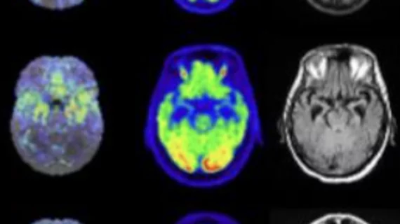
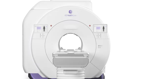
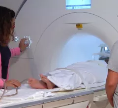
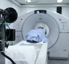
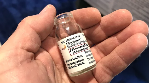
Magnetic resonance imaging (MRI) is the gold standard imaging modality for soft tissues. It produces detail cross-sectional images of soft tissue and bone anatomy, including muscles, tendons, ligaments, brain and organs, without the use of ionizing radiation. In addition to orthopedic imaging, MRI is also used for heart, brain and breast. MRI uses gadolinium contrast in many exams to highlight tissues and blood vessels, which enhances images and offers better diagnostic quality. It can also be used in conjunction with PET scans. How does MRI work? MR creates images by using powerful magnets to polarize hydrogen atoms in water (the body is made of of more than 80% water) so they face in one direction. A radiofrequency pulse is then used to ping these atoms, causing them to wobble, or resonate. The MRI coils detect this and computers can assemble images from the signals. Basic MRI scans will focus on the resonance of fat and water in two different sequences, which highlight and contrast different features in the anatomy.

liver cancer

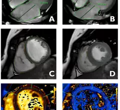
Measurements from functional MRI scans offer non-invasive boost in liver disease assessment

By using a combination of functional liver imaging scores (FLIS) derived from gadoxetic acid (GA)-enhanced MRI and splenic diameter measured on MRI, the experts were able to non-invasively assess liver impairment.

New imaging biomarkers emerge for Alzheimer’s and its impairments

Brain MRI of the choroid plexus, the main supplier of cerebrospinal fluid, can deliver independent biomarkers for Alzheimer’s disease without an assist from clinical tests for amyloid abnormality or neurodegeneration. 

Thumbnail

New 7T MRI scanners could increase radiologists' role in treating neurological conditions

7T MRI brain scans can analyze the locus coeruleusan area difficult to visualize on standard scannersof patients with Parkinson’s and progressive supranuclear palsy to identify those most likely to respond to certain treatment strategies.

AI mammography, prostate imaging algorithms cleared for market

The FDA has OK’d two subsidiaries of Los Angeles-based RadNet to sell medical AI software—one product for diagnosing breast cancer, the other for streamlining MRI prostate reporting workflows. 

Evidence points to optimal MRI sequence for detecting insufficiency fractures

Because sacral insufficiency fractures do not always show a fracture line, they can be difficult to diagnose or even misdiagnosed as bone metastasis, which could result in additional treatments like radio-chemotherapy.

Researchers receive fresh $11M to continue cracking post-TBI epilepsy

Structural brain abnormalities detected by MRI are among the risk factors the funded researchers will identify as novel biomarkers.

Thumbnail

Prostate cancer recurrence scoring system yields 'promising' results

An international panel of experts recently developed and validated a reporting assessment scoring system that analyzes the location and extent of prostate cancer recurrence. 

#imagingequipment #cybersecurity #databreaches

Israel’s famously innovative healthcare system struggles to secure imaging devices

In some cases hospital staff kept no watch over outside-looking-in connections between manufacturers and scanners.