Magnetic Resonance Imaging

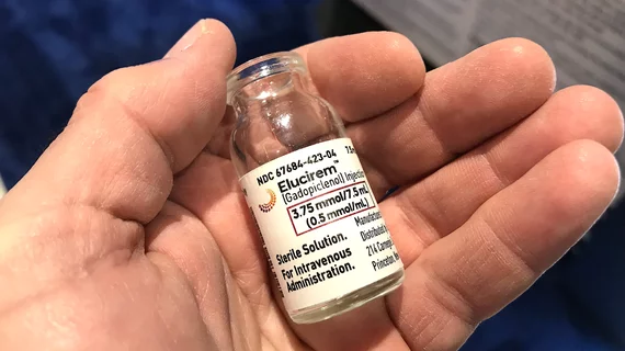
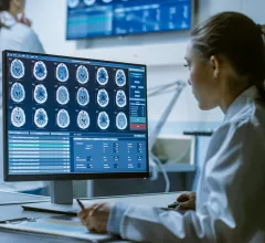
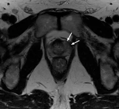
Magnetic resonance imaging (MRI) is the gold standard imaging modality for soft tissues. It produces detail cross-sectional images of soft tissue and bone anatomy, including muscles, tendons, ligaments, brain and organs, without the use of ionizing radiation. In addition to orthopedic imaging, MRI is also used for heart, brain and breast. MRI uses gadolinium contrast in many exams to highlight tissues and blood vessels, which enhances images and offers better diagnostic quality. It can also be used in conjunction with PET scans. How does MRI work? MR creates images by using powerful magnets to polarize hydrogen atoms in water (the body is made of of more than 80% water) so they face in one direction. A radiofrequency pulse is then used to ping these atoms, causing them to wobble, or resonate. The MRI coils detect this and computers can assemble images from the signals. Basic MRI scans will focus on the resonance of fat and water in two different sequences, which highlight and contrast different features in the anatomy.

Thumbnail

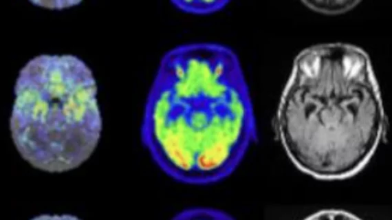
We can now diagnose Alzheimer's using only an MRI and an algorithm

Encouragingly, the MRI scans did not require the latest equipment advances and were conducted on a standard machine that is readily available at most hospitals—a 1.5 Tesla. 

Thumbnail

The benefits of preoperative prostate MRI are clear, so why isn't every man getting one?

Preoperative MRI use increased substantially from 2003 to 2016—from 2.9% to 28.2% to be exact—but the modality remained underutilized in certain regions and among specific populations in the United States.

High-intensity focused ultrasound effectively treats PCA, reduces side effects

Men with prostate cancer can avoid many of the unpleasant side effects of chemo, surgery and radiation therapy when they are treated with high-intensity focused ultrasound instead.

Many clinicians flouting X-ray-first guidelines for ankle imaging

Established clinical guidelines hold that patients presenting with ankle issues should not receive advanced imaging ahead of standard radiography. New research shows a substantial proportion of ordering clinicians sending these patients straight to MRI anyway.

Multimodality imaging turns up serial thromboses following AstraZeneca COVID vaccination

Whole-body imaging reveals clinically undetectable blood clots in patients after their first dose of AstraZeneca's COVID vaccine.

Thumbnail

NIH grants $3.5 million toward researching COVID's neurological impact

The funds will be allocated specifically toward neuroimaging and cognitive and immunological tests to explore if and how mild and asymptomatic SARS-CoV-2 infections have a long-lasting impact on neurocognitive function. 

rib fracture broken ribs

Advanced MSK imaging trio—CT, MR, ultrasound—justified for certain trauma patients upon incident and over time

The comprehensive approach may be warranted for patients whose musculoskeletal injuries include fractures of the costal cartilage. 

Thumbnail

First ever full course of the latest MR-guided radiation therapy treatments completed at Henry Ford Health

The Detroit-based cancer institute achieved this landmark moment using the MRIdian A3i, which is the latest enhancement of the MRIdian system from ViewRay, Inc.