Magnetic Resonance Imaging

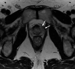
Magnetic resonance imaging (MRI) is the gold standard imaging modality for soft tissues. It produces detail cross-sectional images of soft tissue and bone anatomy, including muscles, tendons, ligaments, brain and organs, without the use of ionizing radiation. In addition to orthopedic imaging, MRI is also used for heart, brain and breast. MRI uses gadolinium contrast in many exams to highlight tissues and blood vessels, which enhances images and offers better diagnostic quality. It can also be used in conjunction with PET scans. How does MRI work? MR creates images by using powerful magnets to polarize hydrogen atoms in water (the body is made of of more than 80% water) so they face in one direction. A radiofrequency pulse is then used to ping these atoms, causing them to wobble, or resonate. The MRI coils detect this and computers can assemble images from the signals. Basic MRI scans will focus on the resonance of fat and water in two different sequences, which highlight and contrast different features in the anatomy.

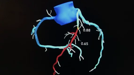
By visualizing metabolism, researchers hope to improve heart disease diagnosis

Hyperpolarized MRI may help identify metabolism problems—which can be an early signal of a heart condition—in real time.

Ultra-high-res MRI Reveals Migraine Brain Changes

7T MRI scans provide a high-res look at how migraines affect the brain

In a new analysis, scheduled to be presented at RSNA 2022 in Chicago, researchers have identified enlarged perivascular spaces in the brains of migraine patients. This may be suggestive of waste clearance issues in the central nervous system.

Thumbnail

Experts ID imaging biomarkers in kids with ADHD

The study's authors explained how MRI results can be used to help identify children with ADHD—and how these findings could be used in the AI algorithms of tomorrow. The team will be presenting its findings at RSNA 2022 in Chicago. 

Canadian radiologists push federal lawmakers to deal with ‘devastating impact’ of long waits for medical imaging

Radiologists from across Canada descended on Parliament Hill in Ottawa earlier this month to voice pressing concerns on behalf of medical-imaging patients and professionals.

Thumbnail

'Significant' brain abnormalities shown to persist 6 months after COVID recovery

The regions that displayed the most significant differentiations in susceptibility values are known to be associated with neurological conditions such as fatigue, insomnia, anxiety, depression, headaches and other cognitive issues—all common complaints among many COVID long haulers. 

How safe is 7T MRI for patients with neurosurgical implants?

These devices have been tested with lower MRI fields, like 1.5T and 3T, but less is known about their compatibility with more powerful 7T equipment. 

Thumbnail

Experts discover brain connectivity patterns unique to children with autism

The findings were also found to be associated with the severity of symptoms in individuals with ASD.

Thumbnail

New lesion measurement better predicts MS progression

Researchers recently presented a new method that considers how already existing lesions grow over time as an indicator of disease progression.