Magnetic Resonance Imaging

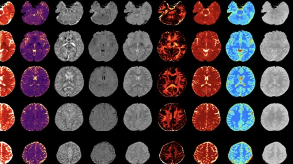
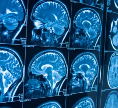
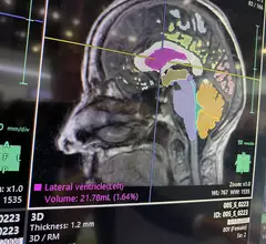
Magnetic resonance imaging (MRI) is the gold standard imaging modality for soft tissues. It produces detail cross-sectional images of soft tissue and bone anatomy, including muscles, tendons, ligaments, brain and organs, without the use of ionizing radiation. In addition to orthopedic imaging, MRI is also used for heart, brain and breast. MRI uses gadolinium contrast in many exams to highlight tissues and blood vessels, which enhances images and offers better diagnostic quality. It can also be used in conjunction with PET scans. How does MRI work? MR creates images by using powerful magnets to polarize hydrogen atoms in water (the body is made of of more than 80% water) so they face in one direction. A radiofrequency pulse is then used to ping these atoms, causing them to wobble, or resonate. The MRI coils detect this and computers can assemble images from the signals. Basic MRI scans will focus on the resonance of fat and water in two different sequences, which highlight and contrast different features in the anatomy.

New data on how pollution affects brain function is 'concerning'

“Air pollution is now recognized as the largest environmental threat to human health and we are increasingly seeing the impacts across all major organ systems," experts explained recently in Environmental Health.

Thumbnail

Advanced PET/MRI protocols boost lung cancer detection

Experts involved in the new research indicate that PET/MRI could have an increasingly prevalent role in lung cancer staging and restaging.

Thumbnail

Prenatal MRI reveals placental damage caused by COVID

In general, any COVID diagnosis raises risks of fetal harm during pregnancy, but fetuses of women who have been diagnosed with a pre-Omicron variant of the virus are most at risk of developmental impairment, according to new research.

Gun goes off during MRI, injuring patient's son

A 40-year-old man was accompanying his mother to her MRI exam when the magnet caused his gun to fire, shooting him in the abdomen.

Thumbnail

MRI and mass spectrometry imaging to play key role in Alzheimer's research

By combining MSI results with whole-brain MRI exams, researchers are hoping to gain insight into the development of Alzheimer’s disease on a molecular level. 

Deep learning slashes real-world MRI scan times

Accelerated MRI with AI image reconstruction nearly halved orthopedic scan times while maintaining or even improving image quality in a newly published prospective study. 

Thumbnail

MRI uncovers clues that could help clinicians better understand schizophrenia

Thanks to functional MRI exams, experts recently gained new insight into mechanisms behind the “loosening of associations” commonly observed in individuals diagnosed with schizophrenia.

#glioblastoma multiforme #GBM #braintumor

Deep learning helps experts take advantage of 'rare' chance to study deadly tumor progression

Researchers from the University of Waterloo and the University of Toronto are collaborating to better understand how glioblastoma multiforme (GBM) advances when it is not treated.